How does the body renew itself? How do cancer cells use the same or similar processes to form tumors and spread throughout the body? How might we use those processes to heal injuries or fight cancer?
A new research program at MIT is tackling fundamental biological questions about normal adult stem cells and their malignant counterparts, cancer stem cells. Launched last spring with support from Fondation MIT, a Swiss philanthropic organization, the MIT Stem Cell Initiative is headed by Jacqueline Lees, the Virginia and D.K. Ludwig Professor of Cancer Research, professor of biology, and associate director of the Koch Institute for Integrative Cancer Research. Other founding members of the initiative are Robert Weinberg, a professor of biology, Whitehead Institute member, and director of the Ludwig Center at MIT; and Omer Yilmaz, an assistant professor of biology.
Rare power
Normal adult stem cells have been defined for more than a half-century. Relatively rare, they are undifferentiated cells within a tissue that divide to produce two daughter cells. One remains in the stem cell state to maintain the stem cell population, a process called self-renewal. The second daughter cell adopts a partially differentiated state, then goes on to divide and differentiate further to yield multiple cell types that form that tissue. In many fully formed adult tissues, normal stem cells divide periodically to replenish or repair the tissue. Importantly, this division is a carefully controlled process to ensure that tissues are restricted to the appropriate size and cell content.
Cancer stem cells are also of long-standing interest and share many similarities with normal adult stem cells. They perform the same division but, rather than differentiating, the additional cells produced by the second daughter cell amass to form the bulk of the tumor. Following surgery or treatment, cancer stem cells can regrow the tumor — and are frequently resistant to chemotherapy — making them especially dangerous. This unique ability of normal and cancer stem cells to both self-renew and form a tissue or tumor is referred to by researchers as “stemness,” and has important implications for biomedical applications.
Because of the key role they play in tissue maintenance and regeneration, normal stem cells hold great promise for use in repairing damaged tissues. Cancer stem cells, correspondingly, are the lifeblood of tumors. Although relatively rare within tumors, they are particularly important because they possess the ability to create tumors and are also chemotherapy-resistant. As a result, cancer stem cells are thought to be responsible for tumor recurrence after remission, and also for the formation of metastases, which account for the majority of cancer-associated deaths. Accordingly, an anti-cancer stem cell therapy that can target and kill cancer stem cells is one of the holy grails of cancer treatment — a means to suppress both tumor recurrence and metastatic disease.
Hiding in plain sight
One of the fundamental challenges to studying normal and cancer stem cells, and to ultimately harnessing that knowledge, is developing the ability to identify, purify, and propagate these cells. This has often proved tricky, as another key similarity between normal and cancer stem cells is that neither is visibly different from other cells in a tissue or tumor. Thus, a major goal in stem cell and cancer stem cell research is finding ways to distinguish these rare specimens from other cells, ideally by identifying unique surface markers that can be used to purify stem cell and cancer stem cell populations and enable their study.
The MIT Stem Cell Initiative is applying new technologies and approaches in pursuit of this goal. More specifically, the program aims to:
- identify the stem cells and cancer stem cells in various tissues and tumor types;
- determine how these cells change during aging (in the case of normal stem cells) or with disease progression (in the case of degenerative conditions and cancer); and
- determine the similarities and differences between normal and cancer stem cells, with the goal of finding vulnerabilities in cancer stem cells that can be viable and specific targets for treatment.
Ultimately, the ability to identify, purify, and establish various populations of stem cells and cancer stem cells could help researchers better understand the biology of these cells, and learn how to utilize them more effectively in regenerative medicine applications and target them in cancer.
When biology meets technology
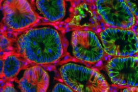
MIT Stem Cell Initiative studies focus on normal and cancer stem cells of epithelial tissues. Epithelia are one of four general tissue types in the body; they line most organs and are where the vast majority of cancers arise. Epithelial cells from different organs share some biological properties, but also have distinct differences reflecting the organ’s specific role and/or environment. In particular, the MIT Stem Cell Initiative has focused on the breast and colon, as these tissues are quite different from each other, yet each constitutes a major portion of cancer incidence.
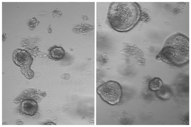
New technologies are enabling the researchers to make significant headway in these investigations, progress that was not feasible just a few years ago. Specifically, they are using a combination of specially cultured cells, sophisticated and highly controllable mouse models of cancer, and single-cell RNA sequencing and computational analysis techniques that are uniquely suited to extracting a great deal of information from the relatively small number of stem cells.
While breast and colon work is ongoing, MIT Stem Cell Initiative members are planning studies of additional tissues and recruiting collaborators for pilot projects. The results of the researchers’ studies will advance understandings of stem cell regulation and may ultimately lead to advances in tissue regeneration and/or cancer analysis and treatment.