Today, the National Institutes of Health (NIH) announced the first round of Brain Research through Advancing Innovative Neurotechnologies (BRAIN) Initiative award recipients, including several MIT interdisciplinary teams.
The BRAIN Initiative, spearheaded by President Obama in April 2013, challenges the nation’s leading scientists to advance our sophisticated understanding of the human mind and discover new ways to treat, prevent, and cure neurological disorders like Alzheimer’s, schizophrenia, autism, and traumatic brain injury.
“The human brain is one of the most complicated structures in the known universe,” said NIH Director Francis Collins. “We have an unprecedented opportunity to develop new technologies that will allow us to map the circuits of the brain, measure activity within those circuits, and understand how their interactions maintain health and modulate human behavior.”
With the NIH Brain Initiative, the scientific community is charged with accelerating the invention of cutting-edge technologies that can produce dynamic images of complex neural circuits and illuminate the interaction of lightning-fast brain cells. These new capabilities are expected to provide greater insights into how brain functionality is linked to behavior, learning, memory, and the underlying mechanisms of debilitating disease.
Exploring perceptual decision-making
One collaborative group of MIT BRAIN Initiative awardees will tap into their diverse expertise to gain a more comprehensive understanding of how cortical circuits process information during memory-guided perceptual decisions.
These investigators are: Mriganka Sur, principal investigator at the Picower Institute for Learning and Memory and the Paul E. Newton Professor of Neuroscience in MIT’s Department of Brain and Cognitive Sciences (BCS); Kwanghun Chung, Picower principal investigator and assistant professor in the Department of Chemical Engineering and the MIT Institute for Medical Engineering and Science (IMES); Ian Wickersham, research scientist at the McGovern Institute for Brain Research and head of MIT’s Genetic Neuroengineering Group; and Emery Brown, professor of computational neuroscience in BCS and the Edward Hood Taplin Professor of Medical Engineering.
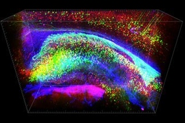
The brain’s cerebral cortex is home to our highest cognitive capacities. Its functionality is enabled by millions of neurons distributed across multiple diverse regions and connected via intricate circuits. Current methods of recording neuron activity involve recording only hundreds of neurons, confined to one region of the cortex.
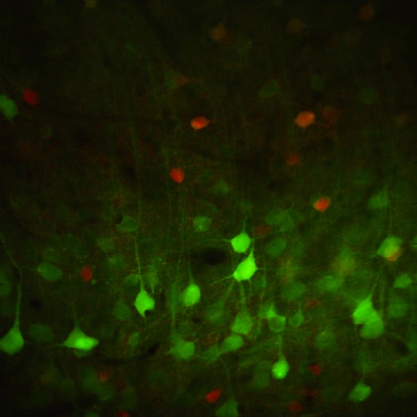
The MIT team will explore the role of multiple regions in mediating behavior. They will accomplish their objectives by developing novel methods of simultaneously recording the activity of thousands of neurons in mice, revealing connections and circuits made by specific neuron types, and employing new statistical methods to analyze massive data sets.
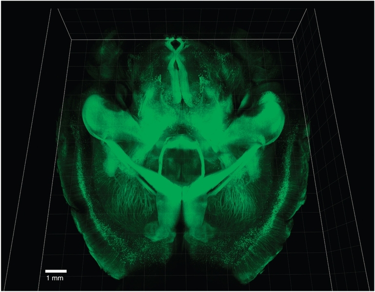
Using innovative optogenetic tools and a method of immunostaining known as CLARITY, they will examine the roles that specific neurons associated with particular regions of the brain play in processing sensory input and choosing actions to complete various parts of a task.
Mriganka Sur received his PhD in electrical engineering from Vanderbilt University. In 1986, he joined the BCS faculty, and in 2000, he became a principal investigator of the Picower Institute. He served as head of BCS from 1997 to 2012, and currently serves as founding director of the Simons Center for the Social Brain. The Sur lab studies cortical function and development with the goal of understanding plasticity and dynamics in networks of the developing and adult visual cortex.
Sur also recently received one of 36 Early Concept Grants for Exploratory Research (EAGER), part of the NSF’s investment in the BRAIN Initiative. Sur will leverage his funding to develop next-generation technologies for recording the activity of massive numbers of cortical neurons across multiple brain regions to reveal the circuits underlying short-term memory. “We intend to develop novel approaches to identify and then stimulate specific neuronal assemblies to determine how they influence behavior and cognition,” said Sur. “These innovative technologies are game changers for neuroscience.”
Kwanghun Chung received his PhD from the Georgia Institute of Technology. He joined the Karl Deisseroth Lab at Stanford University for his postdoctoral training. There, he invented CLARITY, which enables structural and molecular analysis of large-scale intact biological samples. He joined MIT in 2013 and launched a lab devoted to developing and applying novel technologies for integrative and comprehensive understanding of brain function and dysfunction.
Ian Wickersham earned his PhD from the University of California at San Diego. He joined MIT in 2007 as a postdoctoral fellow in the laboratory of Sebastian Seung and subsequently served as a research scientist in Ed Boyden’s lab. In 2013, Wickersham launched the MIT Genetic Neuroengineering Group for developing virus-based genetic tools for neuroscience research.
Wickersham is also the principal investigator on another NIH BRAIN Initiative award for developing systems for nontoxic transsynaptic transgene expression in synaptically connected neurons, in collaboration with Professor Li-Huei Tsai and Assistant Professor Kay Tye, both of the Picower Institute, and Professor Robert Desimone of the McGovern Institute. In addition, Wickersham earned an EAGER award for developing a method of cell-type specific optogenetics in wild-type animals, again in collaboration with Tsai, Tye, and Desimone.
Emery Brown received his MD and PhD in statistics from Harvard University. He is co-director of Harvard-MIT Health Sciences and Technology (HST) and associate director of IMES. Brown’s Neuroscience Statistics Research Lab develops statistical methods and signal-processing algorithms for neuroscience data analysis. He was recently elected to the National Academy of Sciences.
How synapses process information
A second team of MIT BRAIN Initiative awardees will explore how the integration of excitatory and inhibitory inputs (synapses) within a single neuron supports information processing in normally functioning brains — and how it is altered in dysfunctional networks impacted by disease.
This team includes Elly Nedivi, principal investigator at the Picower Institute for Learning and Memory and professor in the MIT departments of Biology and Brain and Cognitive Sciences, and colleague Peter So, MIT mechanical and biological engineering professor.
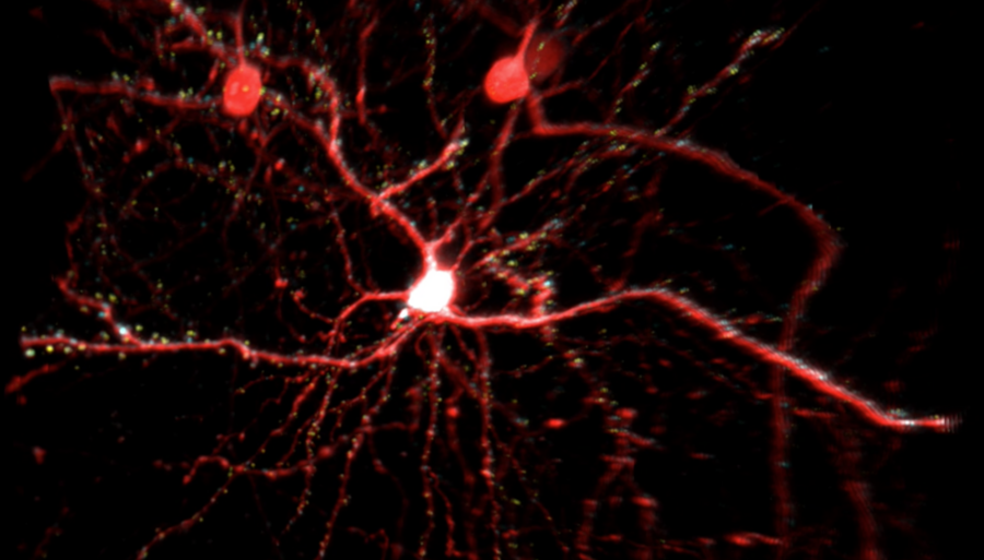
To accomplish their objectives, neurons in a living brain are genetically engineered so that relevant structures are labeled with different color fluorescent proteins allowing high-resolution 3-D mapping of all inputs onto a single neuron. This synaptic map is then used to guide a novel random-access imaging system that can monitor activity at all synaptic sites with millisecond temporal resolution.
The valuable data generated by this research will shed new light on the synaptic transmission and neuronal network interconnectivity that underlie information processing and brain plasticity. The next-generation random access imaging technology proposed by Nedivi and So is expected to enable significantly optimized monitoring of larger, more complex communication networks in animal and human brains.
The Nedivi and So labs have been collaborating for more than a decade to develop methods for monitoring synapses across neurons in the mouse visual cortex. Currently, they are joining forces again to develop next-generation multi-photon microscopy for high-resolution functional brain imaging of synaptic structural dynamics — at the core of the NIH-supported BRAIN Initiative research they will be taking on.
Nedivi earned BS degrees in biology and biochemistry from Hebrew University in Israel and a PhD in neuroscience from Stanford University. She completed postdoctoral work in molecular neurobiology at Israel’s Weizmann Institute. After two years at Cold Spring Harbor Laboratory, Nedivi joined MIT in 1998 as an assistant professor in the Department of Brain and Cognitive Sciences, with a joint appointment in the Department of Biology.
Learning and memory demonstrate the brain’s ability to modify connections in response to input. Plasticity is what allows the brain to constantly adapt to change. The Nedivi lab at the Picower Institute studies the cellular mechanisms that underlie activity-dependent plasticity in the developing and adult brain by examining structural dynamics, and identifying and classifying participating genes and their protein functions.
So earned his BS degrees in physics and mathematics from Harvey Mudd College and his PhD in physics from Princeton University. He continued his postdoctoral research at the Laboratory for Fluorescence Dynamics at University of Illinois. So joined MIT in 1996 as an assistant professor in the Department of Mechanical Engineering. He currently serves as the director of the MIT Laser Biomedical Research Center, an NIH-supported research resource established more than 25 years ago. The So lab creates novel optical imaging, manipulation, and fabrication instrumentation and technologies to address diverse biomedical challenges and drive advances in biology and medicine. So’s group has pioneered numerous optical microscopy and spectroscopy techniques.
The BRAIN Initiative was launched with approximately $100 million in initial investments from the NIH, the National Science Foundation (NSF), and the Defense Advanced Research Projects Agency (DARPA).
Visit the NIH BRAIN Initiative website for more information on the project, or the NSF brain research site for information about NSF support of the BRAIN Initiative.