

Cells are enveloped by a lipid membrane that gives them structure and provides a barrier between the cell and its environment. However, evidence has recently emerged suggesting that these membranes do more than simply provide protection — they also influence the behavior of the protein receptors embedded in them.
A new study from MIT chemists adds further support to that idea. The researchers found that changing the composition of the cell membrane can alter the function of a membrane receptor that promotes proliferation.
Epidermal growth factor receptor (EGFR) can be locked into an overactive state when the cell membrane has a higher than normal concentration of negatively charged lipids, the researchers found. This may help to explain why cancer cells with high levels of those lipids enter a highly proliferative state that allows them to divide uncontrollably.
“The longstanding dogma of what a membrane does is that it’s just a scaffold, an organizational structure. However, there have been increasing observations that suggest that maybe these membrane lipids are actually playing a role in receptor function,” says Gabriela Schlau-Cohen, the Robert T. Haslam and Bradley Dewey Professor of Chemistry at MIT and the senior author of the study.
The findings open up the possibility of discovering new ways to treat tumors by neutralizing the negative charge, which might turn down EGFR signaling, she adds.
Shwetha Srinivasan PhD ’22 is the lead author of the paper, which appears in the journal eLife. Other authors include former MIT postdocs Xingcheng Lin and Raju Regmi, Xuyan Chen PhD ’25, and Bin Zhang, an associate professor of chemistry at MIT.
Receptor dynamics
The EGF receptor, which is found on cells that line body surfaces and organs, is one of many receptors that help control cell growth. Some types of cancer, especially lung cancer and glioblastoma, overexpress the EGF receptor, which can lead to uncontrolled growth.
Like most receptor proteins, EGFR spans the entire cell membrane. Until recently, it has been challenging to study how signals are conveyed across the entire receptor, because of the difficulty of creating membranes that have proteins going all the way through them and then studying both ends of those proteins.
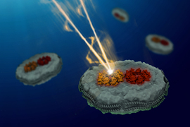
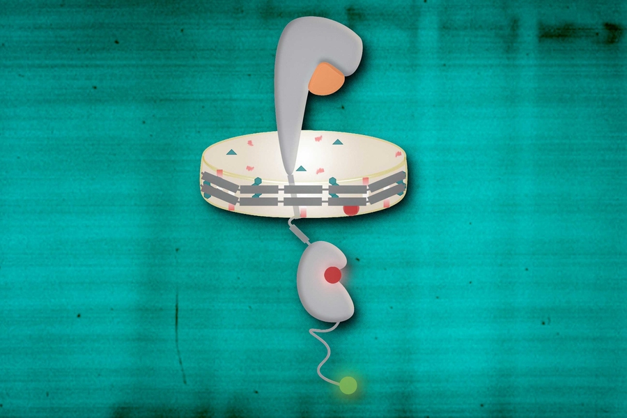
To make it easier to study these signaling processes, Schlau-Cohen’s lab uses nanodiscs, a special type of self-assembling membrane that mimics the cell membrane. When making these discs, the researchers can embed receptors in them, allowing the team to study the function of the full-length receptor.
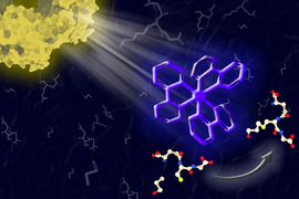
Using a technique called single molecule FRET (fluorescence resonance energy transfer), the researchers can study how the shape of the receptor changes under different conditions. Single molecule FRET allows them to measure the distance between different parts of the protein by labeling them with fluorescent tags and then measuring how fast energy travels between the tags.
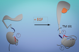
In previous work, Schlau-Cohen and Zhang used single molecule FRET and molecular dynamics simulations to reveal what happens when EGFR binds to EGF. They found that this binding causes the transmembrane section of the receptor to change shape, and that shape-shift triggers the section of the receptor that extends inside the cell to activate cellular machinery that stimulates growth.
Stuck in an overactive state
In the new study, the researchers used a similar approach to investigate how altering the composition of the membrane affects the function of the receptor. First, they explored how elevated levels of negatively charged lipids would affect the cell membrane and EGFR function.
Normally, about 15 percent of the cell membrane is made up of negatively charged lipids. The researchers found that membranes with negatively charged lipids in the range of 15 to 30 percent behaved normally, but if that level reached 60 percent, then the EGFR receptor would become locked into an active state.
In that state, the pro-growth signaling pathway is turned on all the time, even when no EGF is bound to the receptor. Many cancer cells show increased levels of these lipids, and this mechanism could help to explain why those cells are able to grow unchecked, Schlau-Cohen says.
“If the membrane has high levels of negatively charged lipids, then it’s always in that open conformation. It doesn’t matter if ligand is bound or unbound,” she says. “It’s always in the conformation that’s telling the cell to grow, not just when EGF binds.”
The researchers also used this system to explore the role of cholesterol in EGFR function. When the researchers created nanodiscs with elevated cholesterol levels, they found that the membranes became more rigid, and this rigidity suppressed EGFR signaling.
The research was funded by the National Institutes of Health and MIT’s Department of Chemistry.