
By mining data from X-ray images, researchers at MIT, Stanford University, SLAC National Accelerator, and the Toyota Research Institute have made significant new discoveries about the reactivity of lithium iron phosphate, a material used in batteries for electric cars and in other rechargeable batteries.
The new technique has revealed several phenomena that were previously impossible to see, including variations in the rate of lithium intercalation reactions in different regions of a lithium iron phosphate nanoparticle.
The paper’s most significant practical finding — that these variations in reaction rate are correlated with differences in the thickness of the carbon coating on the surface of the particles — could lead to improvements in the efficiency of charging and discharging such batteries.
“What we learned from this study is that it’s the interfaces that really control the dynamics of the battery, especially in today’s modern batteries made from nanoparticles of the active material. That means that our focus should really be on engineering that interface,” says Martin Bazant, the E.G. Roos Professor of Chemical Engineering and a professor of mathematics at MIT, who is the senior author of the study.
Courtesy of the researchers
This approach to discovering the physics behind complex patterns in images could also be used to gain insights into many other materials, not only other types of batteries but also biological systems, such as dividing cells in a developing embryo.
“What I find most exciting about this work is the ability to take images of a system that’s undergoing the formation of some pattern, and learning the principles that govern that,” Bazant says.
Hongbo Zhao PhD ’21, a former MIT graduate student who is now a postdoc at Princeton University, is the lead author of the new study, which appears today in Nature. Other authors include Richard Bratz, the Edwin R. Gilliland Professor of Chemical Engineering at MIT; William Chueh, an associate professor of materials science and engineering at Stanford and director of the SLAC-Stanford Battery Center; and Brian Storey, senior director of Energy and Materials at the Toyota Research Institute.
“Until now, we could make these beautiful X-ray movies of battery nanoparticles at work, but it was challenging to measure and understand subtle details of how they function because the movies were so information-rich,” Chueh says. “By applying image learning to these nanoscale movies, we can extract insights that were not previously possible.”
Modeling reaction rates
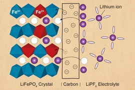
Lithium iron phosphate battery electrodes are made of many tiny particles of lithium iron phosphate, surrounded by an electrolyte solution. A typical particle is about 1 micron in diameter and about 100 nanometers thick. When the battery discharges, lithium ions flow from the electrolyte solution into the material by an electrochemical reaction known as ion intercalation. When the battery charges, the intercalation reaction is reversed, and ions flow in the opposite direction.
“Lithium iron phosphate (LFP) is an important battery material due to low cost, a good safety record, and its use of abundant elements,” Storey says. “We are seeing an increased use of LFP in the EV market, so the timing of this study could not be better.”
Before the current study, Bazant had done a great deal of theoretical modeling of patterns formed by lithium-ion intercalation. Lithium iron phosphate prefers to exist in one of two stable phases: either full of lithium ions or empty. Since 2005, Bazant has been working on mathematical models of this phenomenon, known as phase separation, which generates distinctive patterns of lithium-ion flow driven by intercalation reactions. In 2015, while on sabbatical at Stanford, he began working with Chueh to try to interpret images of lithium iron phosphate particles from scanning transmission X-ray microscopy.
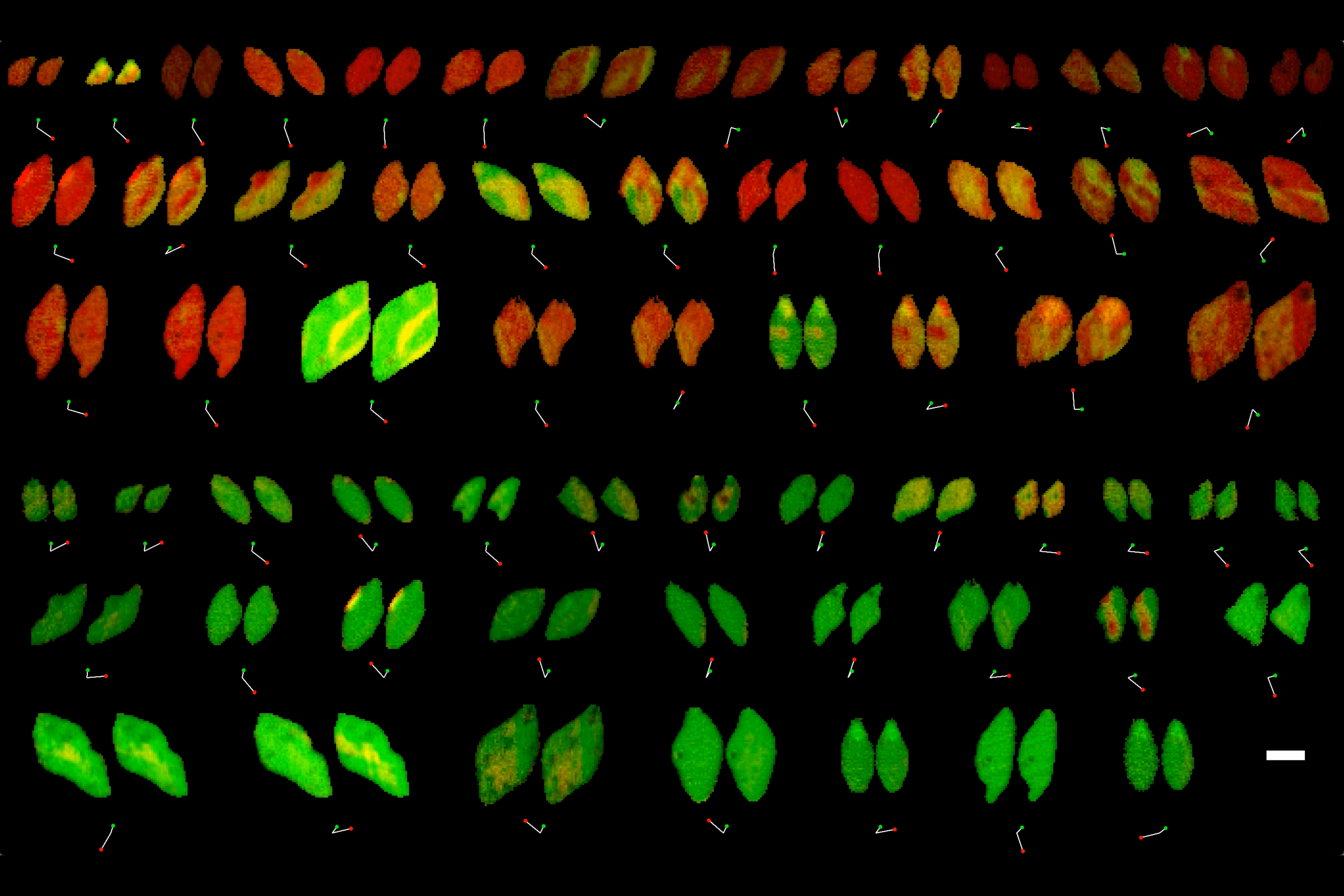
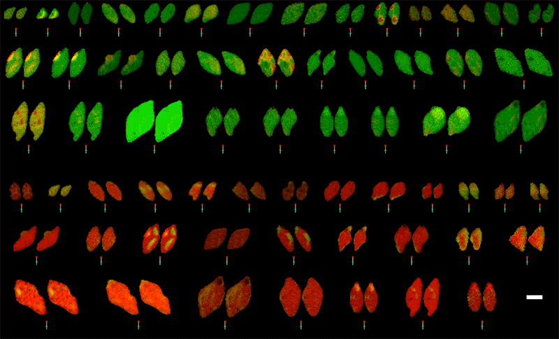
Using this type of microscopy, the researchers can obtain images that reveal the concentration of lithium ions, pixel-by-pixel, at every point in the particle. They can scan the particles several times as the particles charge or discharge, allowing them to create movies of how lithium ions flow in and out of the particles.
In 2017, Bazant and his colleagues at SLAC received funding from the Toyota Research Institute to pursue further studies using this approach, along with other battery-related research projects.
By analyzing X-ray images of 63 lithium iron phosphate particles as they charged and discharged, the researchers found that the movement of lithium ions within the material could be nearly identical to the computer simulations that Bazant had created earlier. Using all 180,000 pixels as measurements, the researchers trained the computational model to produce equations that accurately describe the nonequilibrium thermodynamics and reaction kinetics of the battery material.
“Every little pixel in there is jumping from full to empty, full to empty. And we’re mapping that whole process, using our equations to understand how that’s happening,” Bazant says.
The researchers also found that the patterns of lithium-ion flow that they observed could reveal spatial variations in the rate at which lithium ions are absorbed at each location on the particle surface.
“It was a real surprise to us that we could learn the heterogeneities in the system — in this case, the variations in surface reaction rate — simply by looking at the images,” Bazant says. “There are regions that seem to be fast and others that seem to be slow.”
Furthermore, the researchers showed that these differences in reaction rate were correlated with the thickness of the carbon coating on the surface of the lithium iron phosphate particles. That carbon coating is applied to lithium iron phosphate to help it conduct electricity — otherwise the material would conduct too slowly to be useful as a battery.
“We discovered at the nano scale that variation of the carbon coating thickness directly controls the rate, which is something you could never figure out if you didn't have all of this modeling and image analysis,” Bazant says.
The findings also offer quantitative support for a hypothesis Bazant formulated several years ago: that the performance of lithium iron phosphate electrodes is limited primarily by the rate of coupled ion-electron transfer at the interface between the solid particle and the carbon coating, rather than the rate of lithium-ion diffusion in the solid.
Optimized materials
The results from this study suggest that optimizing the thickness of the carbon layer on the electrode surface could help researchers to design batteries that would work more efficiently, the researchers say.
“This is the first study that's been able to directly attribute a property of the battery material with a physical property of the coating,” Bazant says. “The focus for optimizing and designing batteries should be on controlling reaction kinetics at the interface of the electrolyte and electrode.”
“This publication is the culmination of six years of dedication and collaboration,” Storey says. “This technique allows us to unlock the inner workings of the battery in a way not previously possible. Our next goal is to improve battery design by applying this new understanding.”
In addition to using this type of analysis on other battery materials, Bazant anticipates that it could be useful for studying pattern formation in other chemical and biological systems.
This work was supported by the Toyota Research Institute through the Accelerated Materials Design and Discovery program.