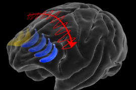
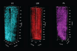
When it comes to processing vision, the brain is full of noise. Information moves from the eyes through many connections in the brain. Ideally, the same image would be reliably represented the same way each time, but instead different groups of cells in the visual cortex can become stimulated by the same scenes. So how does the brain ultimately ensure fidelity in processing what we see? A team of neuroscientists in the Picower Institute for Learning and Memory at MIT found out by watching the brains of mice while they watched movies.
What the researchers discovered is that while groups of “excitatory” neurons respond when images appear, thereby representing them in the visual cortex, activity among two types of “inhibitory” neurons combines in a neatly arranged circuit behind the scenes to enforce the needed reliability. The researchers were not only able to see and analyze the patterns of these neurons working, but once they learned how the circuit operated they also took control of the inhibitory cells to directly manipulate how consistently excitatory cells represented images.
“The question of reliability is hugely important for information processing and particularly for representation — in making vision valid and reliable,” says Mriganka Sur, the Newton Professor of Neuroscience in MIT’s Department of Brain and Cognitive Sciences and senior author of the new study in the Journal of Neuroscience. “The same neurons should be firing in the same way when I look at something, so that the next time and every time I look at it, it’s represented consistently.”
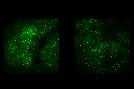
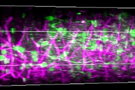
Research scientist Murat Yildirim and former graduate student Rajeev Rikhye led the study, which required a number of technical feats. To watch hundreds of excitatory neurons and two different inhibitory neurons at work, for instance, they needed to engineer them to flash in distinct colors under different colors of laser light in their two-photon microscope. Taking control of the cells using a technology called “optogenetics” required adding even more genetic manipulations and laser colors. Moreover, to make sense of the cellular activity they were observing, the researchers created a computer model of the tripartite circuit.
“It was exciting to be able to combine all these experimental elements, including multiple different laser colors, to be able to answer this question,” Yildirim says.
Reliable representation
The team’s main observation was that as mice watched the same movies repeatedly, the reliability of representation among excitatory cells varied along with the activity levels of two different inhibitory neurons. When reliability was low, activity among parvalbumin-expressing (PV) inhibitory neurons was high and activity among somatostatin-expressing (SST) neurons was low. When reliability was high, PV activity was low and SST activity was high. They also saw that SST activity followed PV activity in time after excitatory activity had become unreliable.
PV neurons inhibit excitatory activity to control their gain, Sur says. If they didn’t, excitatory neurons would become saturated amid a flood of incoming images and fail to keep up. But this gain suppression apparently comes at the cost of making representation of the same scenes by the same cells less reliable, the study suggests. SST neurons meanwhile, can inhibit the activity of PV neurons. In the team’s computer model, they represented the tripartite circuit and were able to see that SST neuron inhibition of PV neurons kicks in when excitatory activity has become unreliable.
"This was highly innovative research for Rajeev's doctoral thesis," Sur says.
The team was able to directly show this dynamic by taking control of PV and SST cells with optogenetics. For instance, when they increased SST activity they could make unreliable neuron activity more reliable. And when they increased PV activity, they could ruin reliability if it was present.
Importantly, though, they also saw that SST neurons cannot enforce reliability without PV cells being in the mix. They hypothesize that this cooperation is required because of differences in how SST and PV cells inhibit excitatory cells. SST cells only inhibit excitatory cell activity via connections, or “synapses,” on the spiny tendrils called dendrites that extend far out from the cell body, or “soma.” PV cells inhibit activity at the excitatory cell body itself. The key to improving reliability is enabling more activity at the cell body. To do that, SST neurons must therefore inhibit the inhibition provided by PV cells. Meanwhile, suppressing activity in the dendrites might reduce noise coming into the excitatory cell from synapses with other neurons.
“We demonstrate that the responsibility of modulating response reliability does not lie exclusively with one neuronal subtype,” the authors wrote in the study. “Instead, it is the co-operative dynamics between SST and PV [neurons] which is important for controlling the temporal fidelity of sensory processing. A potential biophysical function of the SSTàPV circuit may be to maximize the signal-to-noise ratio of excitatory neurons by minimizing noise in the synaptic inputs and maximizing spiking at the soma.”
Sur notes that the activity of SST neurons is not just modulated by automatic feedback from within this circuit. They might also be controlled by “top-down” inputs from other brain regions. For instance, if we realize a particular image or scene is important, we can volitionally concentrate on it. That may be implemented by signaling SST neurons to enforce greater reliability in excitatory cell activity.
In addition to Sur, Yildirim, and Rikhye, the paper’s other authors are Ming Hu and Vincent Breton-Provencher.
The National Eye Institute, The National Institute of Biomedical Imaging and Bioengineering of the National Institutes of Health, and the JPB Foundation funded the study.

