To focus on what’s new, we disregard what’s not. A new study by researchers at MIT’s Picower Institute for Learning and Memory substantially advances understanding of how a mammalian brain enables this “visual recognition memory.”
Dismissing the things in a scene that have proven to be of no consequence is an essential function because it allows animals and people to quickly recognize the new things that need to be assessed, says Mark Bear, Picower Professor in the Department of Brain and Cognitive Sciences and senior author of the study in the Journal of Neuroscience.
“Everyone’s appropriate behavioral response to an unexpected stimulus is to devote attentional resources to that,” Bear says. “Maybe it means danger. Maybe it means food. But if you learn that this once-novel stimulus isn’t anything of significance, it is super adaptive to no longer pay attention to it. It’s absolutely essential for normal brain function that we’re able to make a quick determination of whether a stimulus is novel or not.”
People with schizophrenia and some autism spectrum disorders appear to struggle with this capability, Bear notes.
In 2006, Bear’s lab discovered the first sign of visual recognition memory. Researchers detected a strong pattern of increasing electrical activity in the visual cortex as mice became familiar with an image on a screen. Subsequent research showed that this increase in electrophysiological response, dubbed SRP, or “stimulus selective response plasticity,” correlated strongly with “habituation,” or the behavioral loss of interest in exploring the increasingly familiar stimulus.
Since then, the lab has been working in mice to understand exactly how these phenomena emerge. Their research has shown that a well-known mechanism of learning and memory called “LTP,” a strengthening of neural connections amid frequent activity, is involved, but that this mechanism alone cannot produce visual recognition memory. Inhibitory neurons called parvalbumin (PV) expressing neurons also appear to be crucial parts of the circuit. PV neurons are known to produce high-frequency gamma rhythms in the cortex.
In the new study led by graduate students Dustin Hayden and Daniel Montgomery, Bear’s lab shows that as novel visual patterns become familiar, the transition is marked by stark changes in the visual cortex. Gamma rhythms give way to lower-frequency beta rhythms and the activity of PV neurons dies out in favor of a rise in activity by inhibitory somatostatin (SOM) expressing neurons.
The study, Bear says, therefore provides an externally measurable indicator of the transition from novel to familiar — the brain rhythm shift. It also offers a new hypothesis for how visual recognition memory is enforced: PV activity, which initially inhibits the SRP electrical response, eventually itself becomes inhibited by SOM activity.
Bear’s lab is working with Boston Children’s Hospital researcher Chuck Nelson to determine if aberrations in SRP, such as this frequency transition, can be used as an early biomarker of autism spectrum disorders.
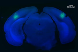
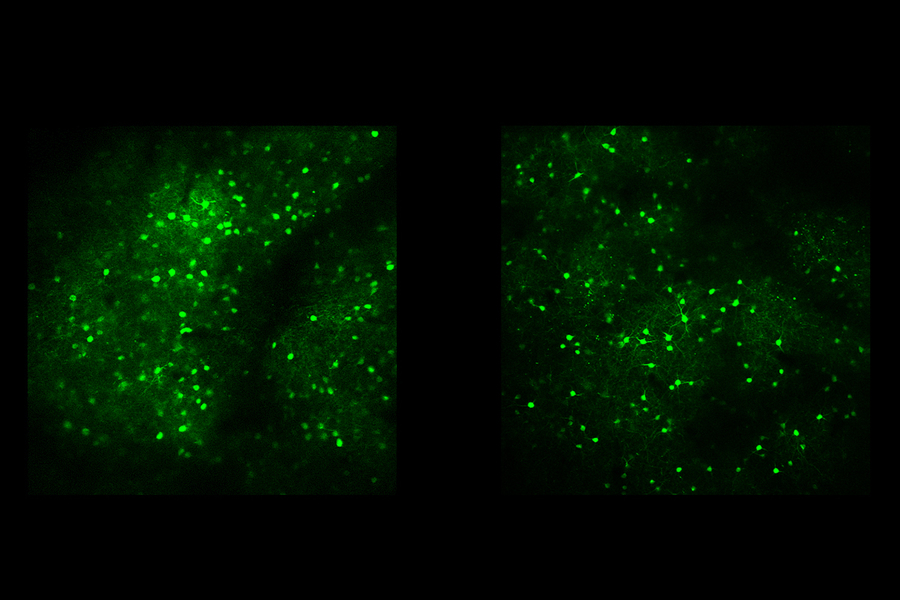
In the new study, the researchers showed mice the same simple image repeatedly over the course of several days. All the while, they measured the SRP electrical response in the mice as well as neural rhythms. In parallel experiments, they engineered some mice so that their PV or SOM neurons would flash brightly when active. Then as mice watched the image, the scientists could watch for those flashes using a “two-photon” microscope.
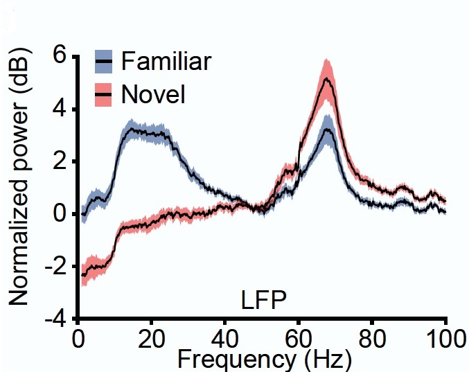
On Day One, when the image was novel, the spectrum of rhythms in the visual cortex was dominated by higher-frequency gamma readings. As the days went on, gamma power diminished, replaced by a steady increase in low-frequency beta power. To ensure this wasn’t an unrelated transition, on Day Seven the scientists presented a new image and the familiar one. When the mice saw the new one, they again exhibited a gamma frequency dominated pattern. When they saw the same old original image, the visual cortex reproduced the pattern of increased beta power.
In a subsequent data analysis, the researchers found that the decline in gamma power and increase in beta power correlated significantly with the SRP growth of electrical activity, suggesting that they are indeed linked.
“These data are compatible with the hypothesis that the same underlying biology is responsible for both manifestations of SRP,” the researchers wrote.
The two-photon microscope experiments revealed that underlying biology difference. PV neurons responded strongly to images when they were novel, but that activity became replaced by increasing SOM activity over several days as the image became familiar.
SOM neurons are believed to suppress PV neurons, Bear says, but to prove that SOM inhibition of PV accounts for visual recognition memory, the lab still needs to perform more experiments. Using optogenetics, in which they can engineer neurons to be controlled with different colors of light, they can manipulate SOM neurons to see if turning them off causes mice to regard repeated images as forever new, or turning them on causes mice to immediately dismiss novel images as passe.
Sam Cooke, a former member of the Bear lab who is now a senior lecturer at Kings College London, is a co-author of the study.
The National Eye Institute, The JPB Foundation, and the National Science Foundation provided funding for the study.