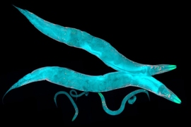
For a nematode worm, a big lawn of the bacteria that it eats is a great place for it to disperse its eggs so that each hatchling can emerge into a nutritive environment. That’s why when a worm speedily roams about a food patch, it methodically lays its eggs as it goes. A new study by neuroscientists at MIT’s Picower Institute for Learning and Memory investigates this example of action coordination — where egg-laying is coupled to the animal’s roaming — to demonstrate how a nervous system coordinates distinct behavioral outputs. That’s a challenge many organisms face, albeit in different ways, during daily life.
“All animals display a remarkable ability to coordinate their diverse motor programs, but the mechanisms within the brain that allow for this coordination are poorly understood,” note the scientists, including Steven Flavell, Lister Brothers Career Development Assistant Professor in MIT’s Department of Brain and Cognitive Sciences.
Flavell lab members Nathan Cermak, Stephanie Yu, and Rebekah Clark were co-lead authors of the study published this month in eLife.
A new imaging platform
To learn how animals coordinate their motor programs, Flavell’s team invented a new microscopy platform capable of taking sharp, high-frame-rate videos of nematodes for hours or days on end. Guided by custom software, the scope automatically tracks the worms, allowing the researchers to compile information about each animal’s behavior. The team also wrote machine vision software to automatically extract information about each of the C. elegans motor programs — locomotion, feeding, egg-laying, and more — from these videos, yielding a near-comprehensive picture of each animal’s behavioral outputs. Flavell said the scope parts cost about $3,000 and can be assembled in a day or two using the team’s online tutorial. They have posted that and the system’s software online for free. The affordability and flexibility of these microscopes should allow them to be useful for many different applications in the biological sciences.
By using this system and then analyzing the data, Flavell’s team was able to identify for the first time a number of patterns of nematode behavior that involve the coordination of multiple motor actions. Flavell said one insight yielded by the system and the subsequent analysis is that the intensely studied nematodes, known scientifically as C. elegans, have more distinct behavioral states than generally assumed. For example, the study finds that the behavioral state known as “dwelling,” previously defined based on the animal staying put, actually consists of multiple different sub-states that could be readily identified using this new imaging approach.
Behaviors coordinated by dopamine
But one of the most pronounced new behavioral patterns that emerged from the analyses was the observation that worms lay many more eggs while roaming on a food lawn than they do while dwelling. This likely allows animals to thoroughly disperse their eggs across a nutritive environment. The two motor circuits that control locomotion and egg-laying in this animal had been carefully defined by previous work. So, based on their new observation, Flavell’s team decided to investigate how the worm’s nervous system couples locomotion and egg-laying together. It turned out to hinge on the neurotransmitter dopamine, which is abundant in all animals, including humans.
They started out by knocking out genes for various neurotransmitters and other brain-modulating molecules. Many of those candidates, such as serotonin, affected the animal’s behavior in important ways, but did not disrupt this coupling of roaming and egg laying. It was only when the team knocked out a gene called cat-2, which is needed for dopamine production, that the worms no longer increased their egg laying while roaming. Notably, it didn’t affect the pace of egg laying while dwelling, suggesting that the worms without dopamine were still capable of laying eggs normally while engaged in other behavioral states.
The team further confirmed the role of dopamine by taking direct control of dopamine-producing cells using optogenetics, a technology that allows neuron activity to be turned on or off with flashes of light. In these experiments, they learned that acutely shutting down the dopaminergic neurons reduced egg-laying only while animals were in the roaming state, but activating these neurons could drive the animals to start laying eggs, even under circumstances when the pace of egg-laying is normally low.
Next, the team wanted to know where the dopamine that triggers this coordinated response emerges, and when. They engineered worms so that their neurons would glow when they became electrically active, an indication provided by a surge of calcium ions. From those flashes they saw that a particular dopamine-producing neuron called PDE stood out as being especially active as worms roamed across a food lawn, and their activity fluctuated in association with the worms’ motion. It peaked, they saw, just before the worm assumed the posture that precipitates egg laying, but only when the worms were crawling along a bacterial food source. Notably, the neuron has the means — a little hair-like structure called a cilium — to sense food outside the worm’s body. These studies suggested that the PDE neuron integrates the presence of food in the environment with the worm’s own motion, generating an activity pattern that essentially reports how quickly worms are progressing through their nutritive environment. The release of dopamine by this neuron, and potentially others as well, could relay this information to the egg-laying circuit, allowing for coordination between the behaviors.
Flavell’s team also mapped out the neural circuitry downstream of dopamine and found that its effects are mediated by two receptors in the D2 family of dopamine receptors (dop-2 and dop-3). In addition, a set of neurons that utilize the neurotransmitter GABA appear to play a critical role downstream of dopamine release. They hypothesize that the role of dopamine may be to send the signal amid plentiful food and roaming behavior to override GABA’s inhibition of egg laying, allowing this behavior to proceed.
Ultimately, egg laying while roaming was just one example of motor program coupling that the lab chose to dissect. Flavell and co-authors note there are many others, too.
“One thing that excites us about this study is that it’s now easy with this new microscopy platform to simultaneously measure each of the main motor programs generated by this animal. Hopefully, we can start thinking about the full repertoire of behaviors that it generates as a complete, coordinated set,” the scientists say.
The research team notes that recently-developed technologies for whole-brain calcium imaging have opened the possibility of measuring neuronal activity throughout the brains of various animals, including the worm.
“To understand these comprehensive neural imaging datasets, it will be important to consider how they relate to the output of the whole brain: the full repertoire of behavioral outputs that an animal generates” Flavell says.
The paper’s other authors are Yung-Chi Huang and Saba Baskoylu. The National Science Foundation, the National Institutes of Health, the JPB Foundation, and the Brain and Behavior Research Foundation supported the research.

!["[W]e hope that our studies of vps-50 will provide insights into human neuropsychiatric disorders,” H. Robert Horvitz says.](/sites/default/files/styles/news_article__archive/public/images/201603/MIT-BehavorialStates.jpg?itok=KbSrJbQQ)