The human genome contains about 23,000 genes, but only a fraction of those genes are turned on inside a cell at any given time. The complex network of regulatory elements that controls gene expression includes regions of the genome called enhancers, which are often located far from the genes that they regulate.
This distance can make it difficult to map the complex interactions between genes and enhancers. To overcome that, MIT researchers have invented a new technique that allows them to observe the timing of gene and enhancer activation in a cell. When a gene is turned on around the same time as a particular enhancer, it strongly suggests the enhancer is controlling that gene.
Learning more about which enhancers control which genes, in different types of cells, could help researchers identify potential drug targets for genetic disorders. Genomic studies have identified mutations in many non-protein-coding regions that are linked to a variety of diseases. Could these be unknown enhancers?
“When people start using genetic technology to identify regions of chromosomes that have disease information, most of those sites don’t correspond to genes. We suspect they correspond to these enhancers, which can be quite distant from a promoter, so it’s very important to be able to identify these enhancers,” says Phillip Sharp, an MIT Institute Professor Emeritus and member of MIT’s Koch Institute for Integrative Cancer Research.
Sharp is the senior author of the new study, which appears today in Nature. MIT Research Assistant D.B. Jay Mahat is the lead author of the paper.
Hunting for eRNA
Less than 2 percent of the human genome consists of protein-coding genes. The rest of the genome includes many elements that control when and how those genes are expressed. Enhancers, which are thought to turn genes on by coming into physical contact with gene promoter regions through transiently forming a complex, were discovered about 45 years ago.
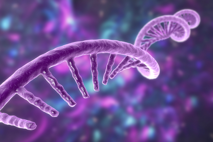
More recently, in 2010, researchers discovered that these enhancers are transcribed into RNA molecules, known as enhancer RNA or eRNA. Scientists suspect that this transcription occurs when the enhancers are actively interacting with their target genes. This raised the possibility that measuring eRNA transcription levels could help researchers determine when an enhancer is active, as well as which genes it’s targeting.
“That information is extraordinarily important in understanding how development occurs, and in understanding how cancers change their regulatory programs and activate processes that lead to de-differentiation and metastatic growth,” Mahat says.
However, this kind of mapping has proven difficult to perform because eRNA is produced in very small quantities and does not last long in the cell. Additionally, eRNA lacks a modification known as a poly-A tail, which is the “hook” that most techniques use to pull RNA out of a cell.
One way to capture eRNA is to add a nucleotide to cells that halts transcription when incorporated into RNA. These nucleotides also contain a tag called biotin that can be used to fish the RNA out of a cell. However, this current technique only works on large pools of cells and doesn’t give information about individual cells.
While brainstorming ideas for new ways to capture eRNA, Mahat and Sharp considered using click chemistry, a technique that can be used to join two molecules together if they are each tagged with “click handles” that can react together.
The researchers designed nucleotides labeled with one click handle, and once these nucleotides are incorporated into growing eRNA strands, the strands can be fished out with a tag containing the complementary handle. This allowed the researchers to capture eRNA and then purify, amplify, and sequence it. Some RNA is lost at each step, but Mahat estimates that they can successfully pull out about 10 percent of the eRNA from a given cell.
Using this technique, the researchers obtained a snapshot of the enhancers and genes that are being actively transcribed at a given time in a cell.
“You want to be able to determine, in every cell, the activation of transcription from regulatory elements and from their corresponding gene. And this has to be done in a single cell because that’s where you can detect synchrony or asynchrony between regulatory elements and genes,” Mahat says.
Timing of gene expression
Demonstrating their technique in mouse embryonic stem cells, the researchers found that they could calculate approximately when a particular region starts to be transcribed, based on the length of the RNA strand and the speed of the polymerase (the enzyme responsible for transcription) — that is, how far the polymerase transcribes per second. This allowed them to determine which genes and enhancers were being transcribed around the same time.
The researchers used this approach to determine the timing of the expression of cell cycle genes in more detail than has previously been possible. They were also able to confirm several sets of known gene-enhancer pairs and generated a list of about 50,000 possible enhancer-gene pairs that they can now try to verify.
Learning which enhancers control which genes would prove valuable in developing new treatments for diseases with a genetic basis. Last year, the U.S. Food and Drug Administration approved the first gene therapy treatment for sickle cell anemia, which works by interfering with an enhancer that results in activation of a fetal globin gene, reducing the production of sickled blood cells.
The MIT team is now applying this approach to other types of cells, with a focus on autoimmune diseases. Working with researchers at Boston Children’s Hospital, they are exploring immune cell mutations that have been linked to lupus, many of which are found in non-coding regions of the genome.
“It’s not clear which genes are affected by these mutations, so we are beginning to tease apart the genes these putative enhancers might be regulating, and in what cell types these enhancers are active,” Mahat says. “This is a tool for creating gene-to-enhancer maps, which are fundamental in understanding the biology, and also a foundation for understanding disease.”
The findings of this study also offer evidence for a theory that Sharp has recently developed, along with MIT professors Richard Young and Arup Chakraborty, that gene transcription is controlled by membraneless droplets known as condensates. These condensates are made of large clusters of enzymes and RNA, which Sharp suggests may include eRNA produced at enhancer sites.
“We picture that the communication between an enhancer and a promoter is a condensate-type, transient structure, and RNA is part of that. This is an important piece of work in building the understanding of how RNAs from enhancers could be active,” he says.
The research was funded by the National Cancer Institute, the National Institutes of Health, and the Emerald Foundation Postdoctoral Transition Award.
