Mirrored chip could enable handheld dark-field microscopes
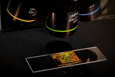
Simple chip powered by quantum dots allows standard microscopes to visualize difficult-to-image biological organisms.
Simple chip powered by quantum dots allows standard microscopes to visualize difficult-to-image biological organisms.
Transmission electron microscope and scanning tunneling microscope offer unique capabilities.
MIT Professor Frances Ross is pioneering new techniques to study materials growth and how structure relates to performance.
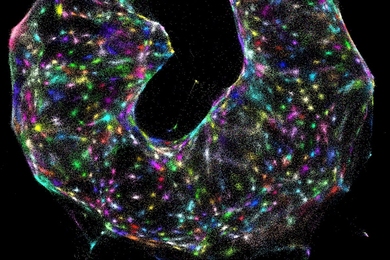
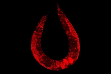
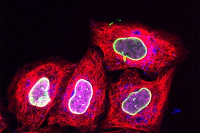
Researchers develop a new microscopy system for creating maps of cells, using chemical reactions to encode spatial information.
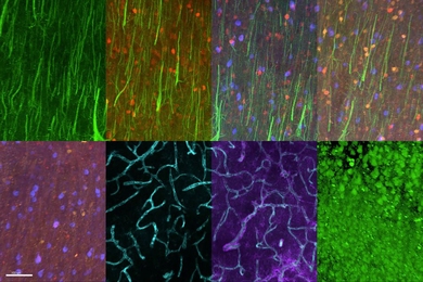
Substantial refinements of three-photon microscopy allow for novel discoveries in neuroscience.
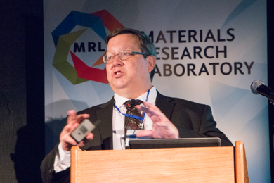
In MIT visit, BP chemist details new X-ray and sample chamber technologies, yielding insights into fighting metal corrosion, improving catalytic reactions, and more.
For a campus that prizes creative risk-taking, Independent Activities Period is a cultural touchstone.
![“…[I]f a fluorescence microscope’s resolution is set at 2 micrometers, our technique can have 300 nanometer resolution — about a sixfold improvement over regular microscopes,” says MIT graduate student Frederick Sangyeon Cho. “The idea is very simple but very powerful and can be useful in many different imaging applications.”](/sites/default/files/styles/term_page__news_article/public/images/201611/MIT-Laser-Particles.jpg?itok=6w5kR_BC)
New imaging technique stimulates particles to emit laser light, could create higher-resolution images.
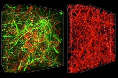
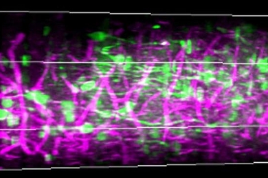
New technique can reveal subcellular details and long-range connections.
Compressing cells allows delivery of new fluorescent tags to track proteins in living cells.
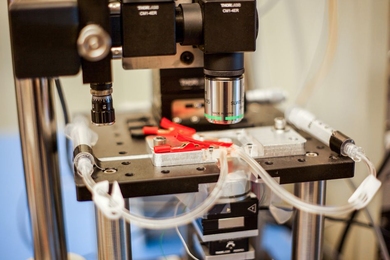
Instrument scans images 2,000 times faster than commercial models.
New technique could contribute to efforts to map the human brain.
MIT physics graduate student James Owen Andrews is developing software to improve dynamic image capture from super-resolution fluorescent microscopes.
Assistant professor of physics probes the formation of enzyme clusters that enable gene copying and protein production in living cells.