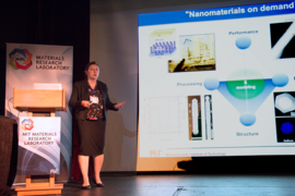
New electron microscopy techniques can help solve corrosion problems that are worth millions of dollars to industrial companies, BP Amoco Chemical Company Senior Research Chemist Matthew Kulzick told the MIT Materials Research Laboratory (MRL) Materials Day Symposium last month.
“Materials science is critical. It’s really material in the financial sense,” Kulzick said. “Solutions demand timely and accurate information. If I’m going to solve a problem, I’ve got to know what’s actually going on, and to do that I need all of these different interrelated tools to be able to go in and find out what’s happening in systems that are important to us.”
New X-ray technologies and sample chambers, he said, are producing stunning images at 20-nanometer scale showing highly localized composition of materials. “The current evolution of tools is spectacular,” he told the audience at the Oct. 10 event.
Beginning in 2003, Kulzick built a new inorganic characterization capability for BP Amoco Chemical, MRL Associate Director Mark Beals said. Kulzick has been working with Nestor J. Zaluzec, a senior scientist at Argonne National Laboratory, as well as with the BP International Center for Advanced Materials, whose partners include the University of Manchester, Imperial College London, the University of Cambridge, and the University of Illinois at Urbana-Champaign.
He outlined advances in imaging technology such as the Pi Steradian Transmission X-ray Detection System developed at the U.S. Department of Energy’s Argonne National Laboratory, and advances in sample holder technology that BP developed collaboratively with Protochips that allow analysis of materials in gas or liquid filled chambers. Microscopic measurements using these holders, or cells, which can include micro-electro-mechanical systems, are called in situ techniques.
“A number of years ago we worked with Protochips, and we modified that holder technology to allow the X-rays coming out of that system to get to the detector,” Kulzick explained.
Images of a palladium and copper-based automotive catalyst from four different generations of energy-dispersive X-ray technology illustrated the evolution from images lacking in detail to a nanoscale compositional image acquired in just 2.5 seconds that shows the location of palladium in the chemical structure.
“So it’s really transformative in understanding what’s happening chemically at the nanoscale,” Kulzick said.
Placing a closed cell filled with hydrogen gas to simulate reduction of the catalyst inside a transmission electron microscope produced images that showed palladium particles remained unaffected while copper particles either migrated toward palladium particles or clustered together with other copper particles.
“We can actually observe the changes that are happening in that localized area under reduction, and this is extremely important if we really want to understand what’s happening,” Kulcizk said. “All of that diversity is occurring in what amounts to roughly a square micron of area on the surface of the material.”
“Just imagine what I could do with this kind of technology with regard to understanding how to activate a catalyst, how to regenerate a catalyst,” he said.
Techniques developed by Professor M. Grace Burke at the University of Manchester in the UK allow observation of chemical changes in a piece of metal over a period of hours such as dissolving a manganese sulfide inclusion from a small piece of stainless steel soaking in water, Kulzick said. “This proved a point for her with regards to corrosion mechanisms that are relevant in the nuclear industry where they worry about what’s initiating crack formation and which she has argued for years that attack of the manganese sulfide by water was one of the underlying mechanisms,” he said.
A significant advance for analyzing organic materials is direct electron capture cameras, Kulzick said.
“One of the problems with bombarding things with electrons is beam damage, so you want to use as little as you can with the right energies,” he added. “The direct electron capture cameras allowed us to reduce that dose.”
For example, Qian Chen, assistant professor of materials science and engineering at the University of Illinois at Urbana-Champaign, has used this enhanced sensitivity and lower dose radiation to a do a series of images at differing tilts to generate a three-dimensional image of a polymer membrane. Computational image analysis becomes important with these 3D structural images. “Without the ability to digitize that material like we’ve done, we would never be able to understand this diversity of structure and make it more rational,” Kulzick said.
Further analysis of the polymer membrane — soaked in a solution of zinc and lead — with Analytical Electron Microscope (AEM) techniques developed by Zaluzec at Argonne National Laboratory revealed that different ions enter into the polymer membrane at different locations. Kulzick said the next step is to understand how ions interact with the membrane structure and how that impacts permeation in the systems. Chen also analyzed the polymer membrane in water inside a graphene cell, Kulzick said, and that work showed swelling of the membrane.
“We hope to put all these pieces together and form a really detailed understanding of how a system like this functions,” he said.


