
Diagnosing cancer with a barcode-inspired test
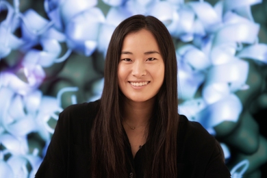
Dana Al-Sulaiman, a recent postdoc with MIT’s Ibn Khaldun Fellowship for Saudi Arabian Women, has developed a cheap, minimally invasive diagnostic test for cancer.
Dana Al-Sulaiman, a recent postdoc with MIT’s Ibn Khaldun Fellowship for Saudi Arabian Women, has developed a cheap, minimally invasive diagnostic test for cancer.
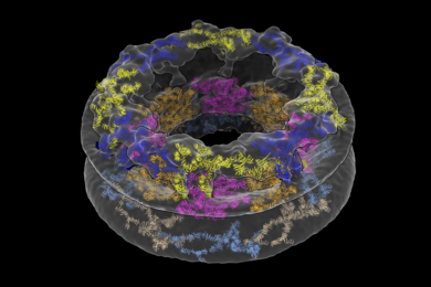
Researchers glean a more complete picture of a structure called the nuclear pore complex by studying it directly inside cells.
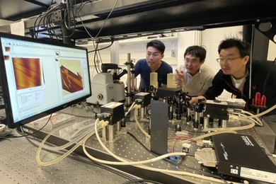
A scattering-type scanning nearfield optical microscope offers advantages to researchers across many disciplines.

MIT researchers train a neural network to predict a “boiling crisis,” with potential applications for cooling computer chips and nuclear reactors.
Researchers could rapidly obtain high-resolution images of blood vessels and neurons within the brain.
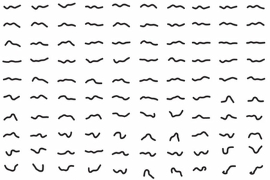
Graduate student Ellen Zhong helped biologists and mathematicians reach across departmental lines to address a longstanding problem in electron microscopy.

Faculty from the departments of Physics and of Nuclear Science and Engineering faculty were selected for the Early Career Research Program.
FIB-SEM is now available to researchers across the Institute for use in characterization, nanofabrication, and rapid prototyping.
Five courses celebrate the nanoscale, highlight technologies in photogrammetry and 360-degree videography.
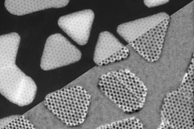
Cutting-edge microscope helps reveal ways to control the electronic properties of atomically thin materials.
Company specializing in atomic force microscopy to advise, collaborate with MIT researchers.
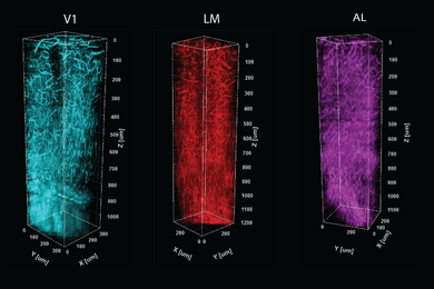
Scientists distinguish brain regions based on what they do, but now have a new way to overlay information about how they are built.
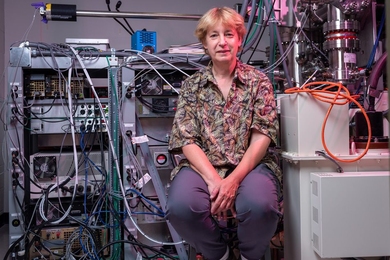
To oversee its new cutting-edge electron microscopy systems, MIT sought out Frances Ross’ industry-honed expertise.
To spy on worms for days on end, Picower Institute scientists invent a new open-source microscopy platform.
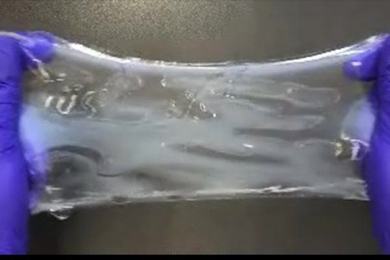
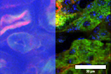
Chemical process called ELAST allows labeling probes to infuse more quickly, and makes samples tough enough for repeated handling.