Within the human brain, neurons perform complex calculations on information they receive. Researchers at MIT have now demonstrated how dendrites — branch-like extensions that protrude from neurons — help to perform those computations.
The researchers found that within a single neuron, different types of dendrites receive input from distinct parts of the brain, and process it in different ways. These differences may help neurons to integrate a variety of inputs and generate an appropriate response, the researchers say.
In the neurons that the researchers examined in this study, it appears that this dendritic processing helps cells to take in visual information and combine it with motor feedback, in a circuit that is involved in navigation and planning movement.
“Our hypothesis is that these neurons have the ability to pick out specific features and landmarks in the visual environment, and combine them with information about running speed, where I’m going, and when I’m going to start, to move toward a goal position,” says Mark Harnett, an associate professor of brain and cognitive sciences, a member of MIT’s McGovern Institute for Brain Research, and the senior author of the study.
Mathieu Lafourcade, a former MIT postdoc, is the lead author of the paper, which appears today in Neuron.
Complex calculations
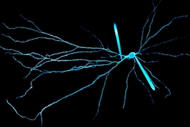
Any given neuron can have dozens of dendrites, which receive synaptic input from other neurons. Neuroscientists have hypothesized that these dendrites can act as compartments that perform their own computations on incoming information before sending the results to the body of the neuron, which integrates all these signals to generate an output.
Previous research has shown that dendrites can amplify incoming signals using specialized proteins called NMDA receptors. These are voltage-sensitive neurotransmitter receptors that are dependent on the activity of other receptors called AMPA receptors. When a dendrite receives many incoming signals through AMPA receptors at the same time, the threshold to activate nearby NMDA receptors is reached, creating an extra burst of current.
This phenomenon, known as supralinearity, is believed to help neurons distinguish between inputs that arrive close together or farther apart in time or space, Harnett says.
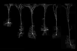
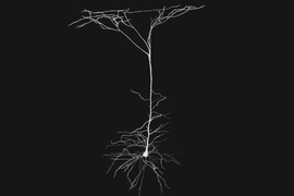
In the new study, the MIT researchers wanted to determine whether different types of inputs are targeted specifically to different types of dendrites, and if so, how that would affect the computations performed by those neurons. They focused on a population of neurons called pyramidal cells, the principal output neurons of the cortex, which have several different types of dendrites. Basal dendrites extend below the body of the neuron, apical oblique dendrites extend from a trunk that travels up from the body, and tuft dendrites are located at the top of the trunk.
Harnett and his colleagues chose a part of the brain called the retrosplenial cortex (RSC) for their studies because it is a good model for association cortex — the type of brain cortex used for complex functions such as planning, communication, and social cognition. The RSC integrates information from many parts of the brain to guide navigation, and pyramidal neurons play a key role in that function.
In a study of mice, the researchers first showed that three different types of input come into pyramidal neurons of the RSC: from the visual cortex into basal dendrites, from the motor cortex into apical oblique dendrites, and from the lateral nuclei of the thalamus, a visual processing area, into tuft dendrites.
“Until now, there hasn't been much mapping of what inputs are going to those dendrites,” Harnett says. “We found that there are some sophisticated wiring rules here, with different inputs going to different dendrites.”
A range of responses
The researchers then measured electrical activity in each of those compartments. They expected that NMDA receptors would show supralinear activity, because this behavior has been demonstrated before in dendrites of pyramidal neurons in both the primary sensory cortex and the hippocampus.
In the basal dendrites, the researchers saw just what they expected: Input coming from the visual cortex provoked supralinear electrical spikes, generated by NMDA receptors. However, just 50 microns away, in the apical oblique dendrites of the same cells, the researchers found no signs of supralinear activity. Instead, input to those dendrites drives a steady linear response. Those dendrites also have a much lower density of NMDA receptors.
“That was shocking, because no one’s ever reported that before,” Harnett says. “What that means is the apical obliques don’t care about the pattern of input. Inputs can be separated in time, or together in time, and it doesn’t matter. It’s just a linear integrator that’s telling the cell how much input it’s getting, without doing any computation on it.”
Those linear inputs likely represent information such as running speed or destination, Harnett says, while the visual information coming into the basal dendrites represents landmarks or other features of the environment. The supralinearity of the basal dendrites allows them to perform more sophisticated types of computation on that visual input, which the researchers hypothesize allows the RSC to flexibly adapt to changes in the visual environment.
In the tuft dendrites, which receive input from the thalamus, it appears that NMDA spikes can be generated, but not very easily. Like the apical oblique dendrites, the tuft dendrites have a low density of NMDA receptors. Harnett’s lab is now studying what happens in all of these different types of dendrites as mice perform navigation tasks.
The research was funded by a Boehringer Ingelheim Fonds PhD Fellowship, the National Institutes of Health, the James W. and Patricia T. Poitras Fund, the Klingenstein-Simons Fellowship Program, a Vallee Scholar Award, and a McKnight Scholar Award.


