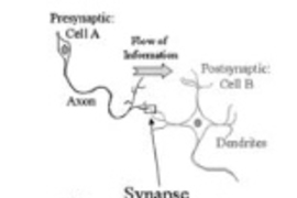
Neuroscientists at MIT’s Picower Institute for Learning and Memory have found that a protein acts like a volume dial for the release of neurotransmitters, the chemicals that neurons release across connections called synapses to stimulate muscles or communicate with other neurons in brain circuits. The findings help explain how synapses work and could better inform understanding of some neurological disorders.
Working in the model of fruit flies, the team determined that the protein Synaptotagmin 7 (SYT7), which is also found in humans and other mammals, constrains the number and availability of neurotransmitter-containing blobs, called vesicles, for release at the synapse. Neurons deploy vesicles to sites called “active zones” to release them across synapses, a process called “vesicle fusion.” When the scientists reduced SYT7, they saw much more neurotransmitter release at synapses. When they increased the protein, neurotransmitter release dropped significantly.
“You can think of this as almost like a radio’s volume dial,” says senior author Troy Littleton, the Menicon Professor of Neuroscience in MIT’s departments of Biology and Brain and Cognitive Sciences. “If a neuron wants to send more signal out, all it has to do is basically reduce the levels of SYT7 protein that it is making. It’s a very elegant way for neurons to turn up or down the amount of output that they are giving.”
The study’s co-lead authors are Zhuo Guan, a research scientist, and Mónica C. Quiñones-Frías, who successfully defended her doctoral thesis on the work May 4. She noted that by acting as that volume dial, the protein could change the nature of a synapse’s activity in a circuit, a property called “synaptic plasticity.”
“Syt7 regulates neurotransmission in a dose-dependent manner and can act as a switch for short-term synaptic plasticity,” Quiñones-Frías says.
Research scientist Yulia Akbergenova is also a co-author of the study published in eLife.
Synaptic surprise
Important as they are, the study’s findings are not ones the team was originally looking for.
For decades, neuroscientists have known that the synaptotagmin protein family plays key roles in synaptic function. In fact, Littleton’s 1993 doctoral dissertation showed that SYT1 promoted a quick release of neurotransmitters when triggered by an influx of calcium ions. But even with SYT1 disabled, synapses could still release neurotransmitters on a slower timeframe. No one has found what promotes that subsequent slower release, but many scientists had pinned their hopes on it being SYT7.
“That’s been something that the whole field, including my lab, has really been searching for,” Littleton says. “So it was a real surprise when we knocked it out and saw just the opposite of what we expected.”
Mutants and microscopes
To study SYT7 the team focused its experiments on synapses in a well-characterized locale: the junction between a fly neuron and muscle. The team not only wanted to see what differences changing the protein’s levels would make in synaptic activity there, but also track how it made those differences.
They changed the amount of SYT7 the neuron could produce by mutating and breeding flies in which the gene was completely eliminated, only one copy could be expressed, or in which the gene was overexpressed, producing more SYT7 than normal. For each of these fly lines they measured the surprising inverse relationship between SYT7 and synaptic transmission.
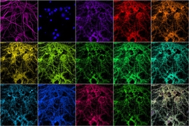
Also, using a technique the lab invented to visually flag neurotransmitter release every time it happens, they mapped how active individual synapses at the neuron-muscle junction were over time. In flies engineered to produce less SYT7 they saw many more synapses with a high propensity for release than they did in normal flies.
Once they confirmed SYT7’s restrictive role, the natural question was how does SYT7 constrain neurotransmitter release. Synapses are very complex, after all, and crucial aspects of SYT7’s role within that machinery had yet to be characterized.
When they compared synapses in normal flies and those missing SYT7 they didn’t see major differences in anatomy or calcium influx that could explain how SYT7 works to limit release.
They then turned their attention to the cycle in which vesicles release their neurotransmitter cargo and are then sent back into the cell to refill with neurotransmitter before rejoining a pool of vesicles ready for redeployment. Their experiments showed that neurons lacking SYT7 didn’t recycle the vesicles differently, but they nevertheless had more vesicles in the readily releasable pool (RRP). Moreover, mutants in which SYT7 was overexpressed substantially limited the vesicles in that pool.
“SYT7 limits release in a dosage-sensitive manner by negatively regulating the number of synaptic vesicles available for fusion and slowing recovery of the RRP following stimulation,” they determined.
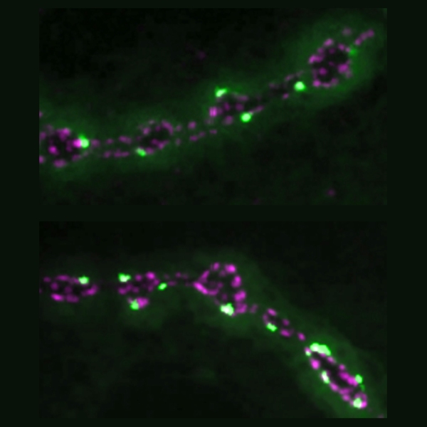
The final step was to track down where SYT7 resides in the synaptic machinery. Under the microscope they were able to pin it down in a network of tubes surrounding, but not within, the active zones. The vantage point is right where other proteins regulating vesicle trafficking also reside, giving SYT7 a clear opportunity to interact with those proteins to regulate the return of vesicles to the active zones.
Implications for disease and plasticity
Understanding more about SYT7’s role at the synapse in mammals could matter in several ways, Littleton says. Two years ago, researchers showed that the protein is reduced in mice harboring a genetic cause of Alzheimer’s disease. And in February another paper showed that patients with bipolar disorder exhibited lower levels of the protein than people who do not have the disorder. Mice with SYT7 knocked out showed some manic and depressive behaviors.
More fundamentally, Littleton and Quiñones-Frías say, is the flexibility or plasticity it can afford. Because SYT7 regulates neurotransmitter release by slowing down the resupply of releasable vesicles, an increase in its levels can transform a synapse from being the kind that sends out large bursts of signal (and therefore transmits more information) early on and then peters out into one that builds up its signal over time. Such distinctions in release timeframe can make important differences in circuit information processing in the brain.
Although the team was able to identify SYT7’s effect at synapses and show key aspects of how it functions, they still hope to determine the exact mechanism that allows the protein to gate vesicle fusion. That work is ongoing.
The National Institutes of Health and the JPB Foundation provided support for the research.