
In addition to responding to electrical and chemical stimuli, many of the body’s neural cells can also respond to mechanical effects, such as pressure or vibration. But these responses have been more difficult for researchers to study, because there has been no easily controllable method for inducing such mechanical stimulation of the cells. Now, researchers at MIT and elsewhere have found a new method for doing just that.
The finding might offer a step toward new kinds of therapeutic treatments, similar to electrically based neurostimulation that has been used to treat Parkinson’s disease and other conditions. Unlike those systems, which require an external wire connection, the new system would be completely contact-free after an initial injection of particles, and could be reactivated at will through an externally applied magnetic field.
The finding is reported in the journal ACS Nano, in a paper by former MIT postdoc Danijela Gregurec, Alexander Senko PhD ’19, Associate Professor Polina Anikeeva, and nine others at MIT, at Boston’s Brigham and Women’s Hospital, and in Spain.
The new method opens a new pathway for the stimulation of nerve cells within the body, which has so far almost entirely relied on either chemical pathways, through the use of pharmaceuticals, or on electrical pathways, which require invasive wires to deliver voltage into the body. This mechanical stimulation, which activates entirely different signaling pathways within the neurons themselves, could provide a significant area of study, the researchers say.
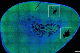
“An interesting thing about the nervous system is that neurons can actually detect forces,” Senko says. “That’s how your sense of touch works, and also your sense of hearing and balance.” The team targeted a particular group of neurons within a structure known as the dorsal root ganglion, which forms an interface between the central and peripheral nervous systems, because these cells are particularly sensitive to mechanical forces.
The applications of the technique could be similar to those being developed in the field of bioelectronic medicines, Senko says, but those require electrodes that are typically much bigger and stiffer than the neurons being stimulated, limiting their precision and sometimes damaging cells.
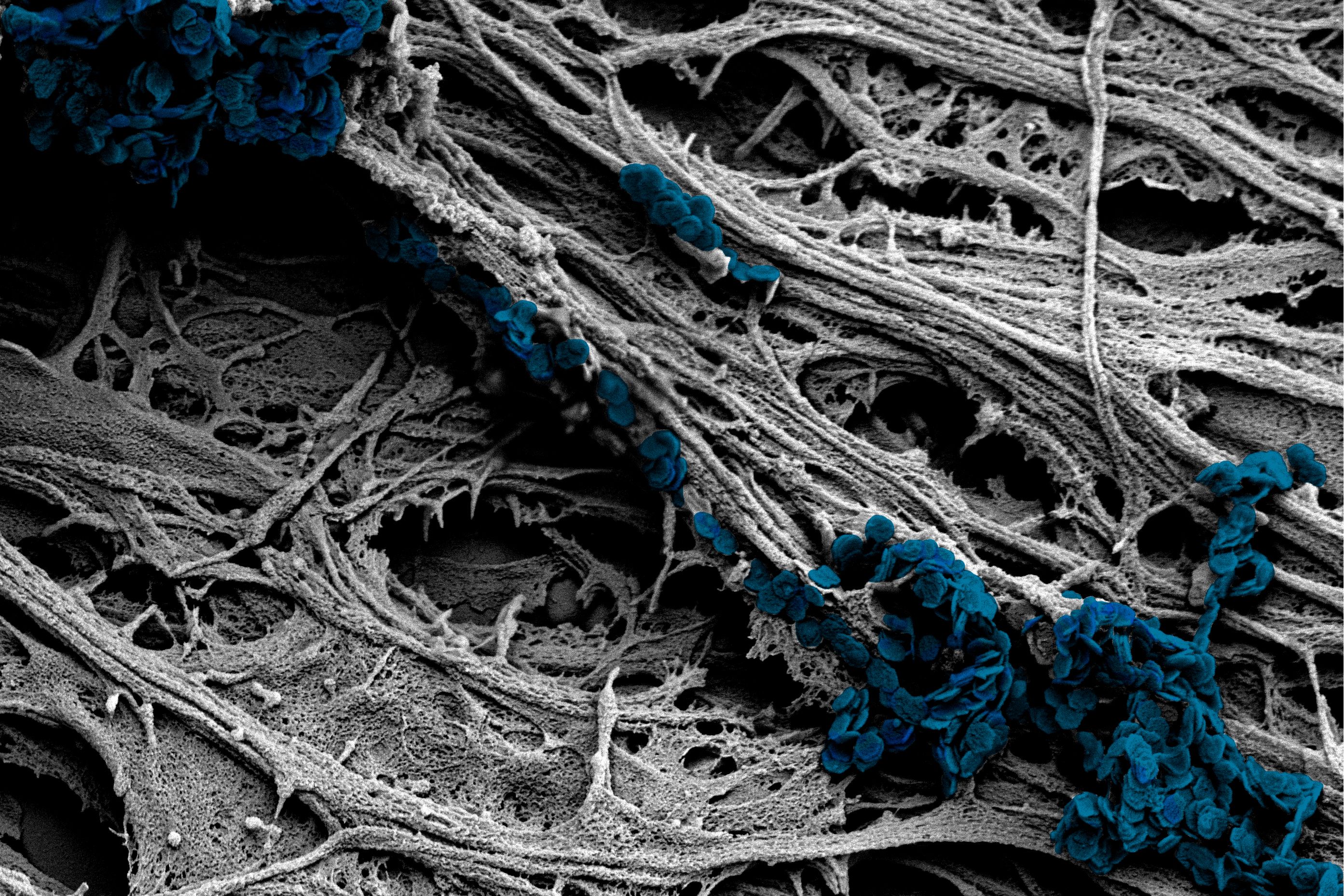
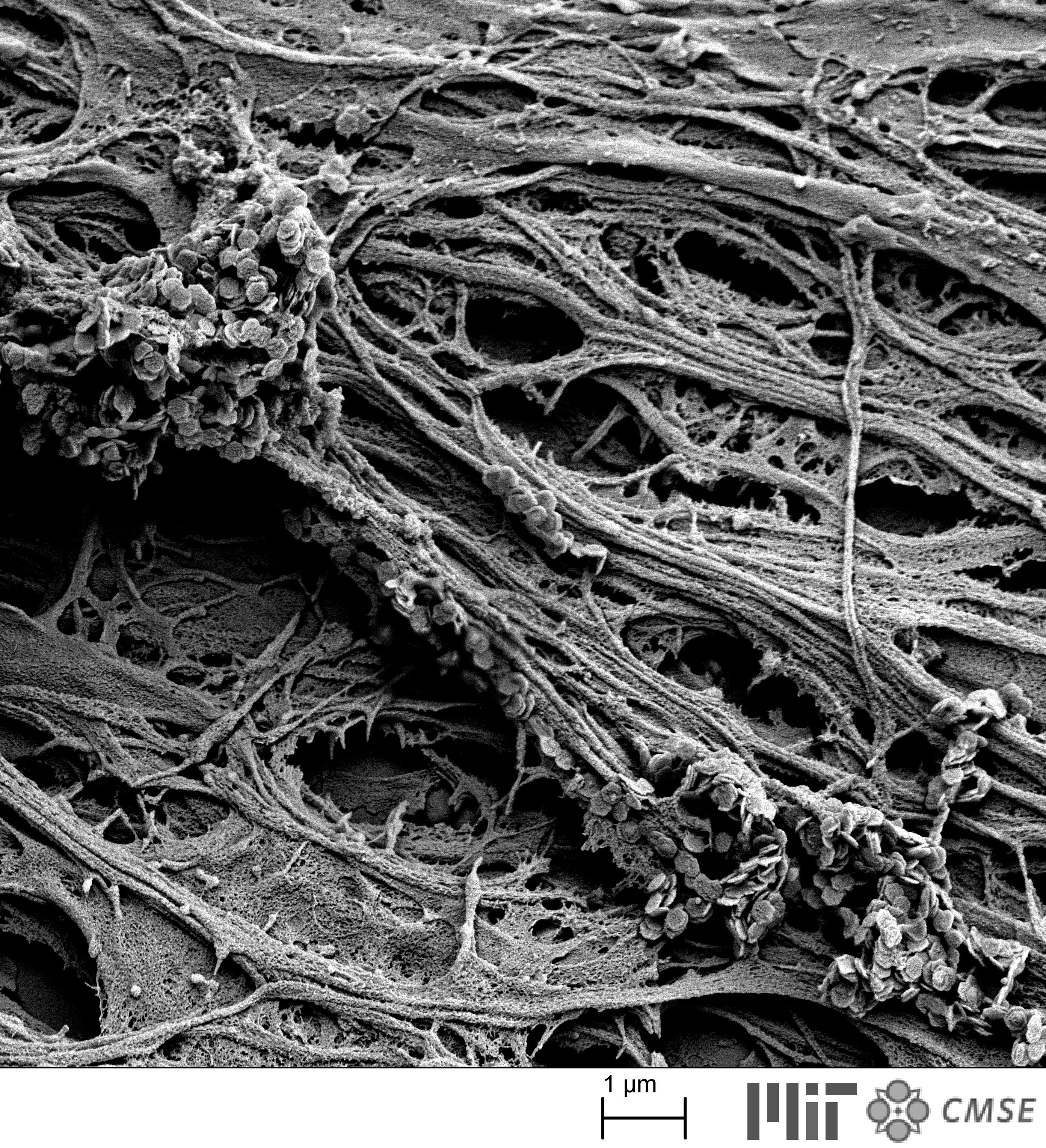
The key to the new process was developing minuscule discs with an unusual magnetic property, which can cause them to start fluttering when subjected to a certain kind of varying magnetic field. Though the particles themselves are only 100 or so nanometers across, roughly a hundredth of the size of the neurons they are trying to stimulate, they can be made and injected in great quantities, so that collectively their effect is strong enough to activate the cell’s pressure receptors. “We made nanoparticles that actually produce forces that cells can detect and respond to,” Senko says.
Anikeeva says that conventional magnetic nanoparticles would have required impractically large magnetic fields to be activated, so finding materials that could provide sufficient force with just moderate magnetic activation was “a very hard problem.” The solution proved to be a new kind of magnetic nanodiscs.
These discs, which are hundreds of nanometers in diameter, contain a vortex configuration of atomic spins when there are no external magnetic fields applied. This makes the particles behave as if they were not magnetic at all, making them exceptionally stable in solutions. When these discs are subjected to a very weak varying magnetic field of a few millitesla, with a low frequency of just several hertz, they switch to a state where the internal spins are all aligned in the disc plane. This allows these nanodiscs to act as levers — wiggling up and down with the direction of the field.
Anikeeva, who is an associate professor in the departments of Materials Science and Engineering and Brain and Cognitive Sciences, says this work combines several disciplines, including new chemistry that led to development of these nanodiscs, along with electromagnetic effects and work on the biology of neurostimulation.
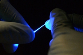
The team first considered using particles of a magnetic metal alloy that could provide the necessary forces, but these were not biocompatible materials, and they were prohibitively expensive. The researchers found a way to use particles made from hematite, a benign iron oxide, which can form the required disc shapes. The hematite was then converted into magnetite, which has the magnetic properties they needed and is known to be benign in the body. This chemical transformation from hematite to magnetite dramatically turns a blood-red tube of particles to jet black.
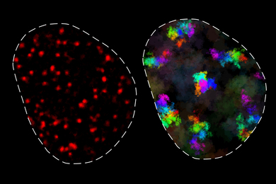
“We had to confirm that these particles indeed supported this really unusual spin state, this vortex,” Gregurec says. They first tried out the newly developed nanoparticles and proved, using holographic imaging systems provided by colleagues in Spain, that the particles really did react as expected, providing the necessary forces to elicit responses from neurons. The results came in late December and “everyone thought that was a Christmas present,” Anikeeva recalls, “when we got our first holograms, and we could really see that what we have theoretically predicted and chemically suspected actually was physically true.”
The work is still in its infancy, she says. “This is a very first demonstration that it is possible to use these particles to transduce large forces to membranes of neurons in order to stimulate them.”
She adds “that opens an entire field of possibilities. … This means that anywhere in the nervous system where cells are sensitive to mechanical forces, and that's essentially any organ, we can now modulate the function of that organ.” That brings science a step closer, she says, to the goal of bioelectronic medicine that can provide stimulation at the level of individual organs or parts of the body, without the need for drugs or electrodes.
The work was supported by the U.S. Defense Advanced Research Projects Agency, the National Institute of Mental Health, the Department of Defense, the Air Force Office of Scientific Research, and the National Defense Science and Engineering Graduate Fellowship.