MIT and Draper researchers have 3-D printed a novel microfluidic device that simulates cancer treatments on biopsied tumor tissue, so clinicians can better examine how individual patients will respond to different therapeutics — before administering a single dose.
Testing cancer treatments today relies mostly on trial and error; patients may undergo multiple time-consuming and hard-to-tolerate therapies in pursuit of one that works. Recent innovations in pharmaceutical development involve growing artificial tumors to test drugs on specific cancer types. But these models take weeks to grow and don’t account for an individual patient’s biological makeup, which can affect treatment efficacy.
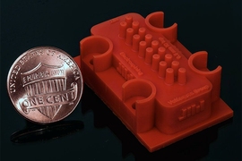
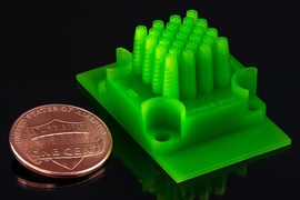
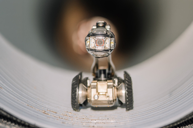
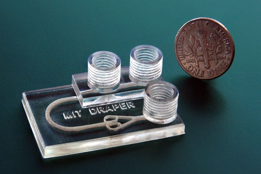
The researchers’ device, which can be printed in about one hour, is a chip slightly larger than a quarter, with three cylindrical “chimneys” rising from the surface. These are ports used to input and drain fluids, as well as remove unwanted air bubbles. Biopsied tumor fragments are placed in a chamber connected to a network of channels that deliver fluids — containing, for instance, immunotherapy agents or immune cells — to the tissue. Clinicians can then use various imaging techniques to see how the tissue responds to the drugs.
A key feature was using a new biocompatible resin — traditionally used for dental applications — that can support long-term survival of biopsied tissue. Although previous 3-D-printed microfluidics have held promise for drug testing, chemicals in their resin usually kill cells quickly. The researchers captured fluorescence microscopy images that show their device, called a tumor analysis platform (TAP), kept more than 90 percent of the tumor tissue alive for at least 72 hours, and potentially much longer.
Because the 3-D printed device is easy and cheap to fabricate, it could be rapidly implemented into clinical settings, the researchers say. Doctors could, for instance, print out a multiplexed device that could support multiple tumor samples in parallel, to enable modeling of the interactions between tumor fragments and many different drugs, simultaneously, for a single patient.
“People anywhere in the world could print our design. You can envision a future where your doctor will have a 3-D printer and can print out the devices as needed,” says Luis Fernando Velásquez-García, a researcher in the Microsystems Technology Laboratories and co-author on a paper describing the device, which appears in the December issue of the Journal of Microelectromechanical Systems. “If someone has cancer, you can take a bit of tissue in our device, and keep the tumor alive, to run multiple tests in parallel and figure out what would work best with the patient’s biological makeup. And then implement that treatment in the patient.”
A promising application is testing immunotherapy, a new treatment method using certain drugs to rev up a patient’s immune system to help it fight cancer. (This year’s Nobel Prize in physiology or medicine was awarded to two immunotherapy researchers who designed drugs that block certain proteins from preventing the immune system from attacking cancer cells.) The researchers’ device could help doctors better identify treatments to which an individual is likely to respond.
“Immunotherapy treatments have been specifically developed to target molecular markers found on the surface of cancer cells. This helps to ensure that the treatment elicits an attack on the cancer directly while limiting negative impacts on healthy tissue. However, every individual’s cancer expresses a unique array of surface molecules — as such, it can be difficult to predict who will respond to which treatment. Our device uses the actual tissue of the person, so is a perfect fit for immunotherapy,” says first author Ashley Beckwith SM ’18, a graduate researcher in Velásquez-García’s research group.
Co-author on the paper is Jeffrey T. Borenstein, a researcher at Draper, where he leads its program in immuno-oncology. “A key challenge in cancer research has been the development of tumor microenvironments that simulate mechanisms of cancer progression and the tumor-killing effects of novel therapeutics,” Borenstein says. “Through this collaboration with Luis and the MTL, we are able to benefit from their great expertise in additive manufacturing technologies and materials science for extremely rapid design cycles in building and testing these systems.”
Supporting cells
Microfluidics devices are traditionally manufactured via micromolding, using a rubberlike material called polydimethylsiloxane (PDMS). This technique, however, was not suitable for creating the three-dimensional network of features — such as carefully sized fluid channels — that mimic cancer treatments on living cells. Instead, the researchers turned to 3-D printing to craft a fine-featured device “monolithically” — meaning printing an object all in one go, without the need to assemble separate parts.
The heart of the device is its resin. After experimenting with numerous resins over several months, the researchers landed finally on Pro3dure GR-10, which is primarily used to make mouthguards that protect against teeth grinding. The material is nearly as transparent as glass, has barely any surface defects, and can be printed in very high resolution. And, importantly, as the researchers determined, it does not negatively impact cell survival.
The team subjected the resin to a 96-hour cytotoxicity test, an assay that exposes cells to the printed material and measures how toxic that material is to the cells. After the 96 hours, the cells in the material were still kicking. “When you print some of these other resin materials, they emit chemicals that mess with cells and kill them. But this doesn’t do that,” Velasquez-Garcia says. “To the best of my knowledge, there’s no other printable material that comes close to this degree of inertness. It’s as if the material isn’t there.”
Setting traps
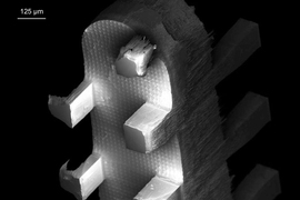
Two other key innovations on the device are the “bubble trap” and a “tumor trap.” Flowing fluids into such a device creates bubbles that can disrupt the experiment or burst, releasing air that destroys tumor tissue.
To fix that, the researchers created a bubble trap, a stout “chimney” rising from the fluid channel into a threaded port through which air escapes. Fluid — including various media, fluorescent markers, or lymphocytes — gets injected into an inlet port adjacent to the trap. The fluid enters through the inlet port and flows past the trap, where any bubbles in the fluid rise up through the threaded port and out of the device. Fluid is then routed around a small U-turn into the tumor’s chamber, where it flows through and around the tumor fragment.
This tumor-trapping chamber sits at the intersection of the larger inlet channel and four smaller outlet channels. Tumor fragments, less than 1 millimeter across, are injected into the inlet channel via the bubble trap, which helps remove bubbles introduced when loading. As fluid flows through the device from the inlet port, the tumor is guided downstream to the tumor trap, where the fragment gets caught. The fluid continues traveling along the outlet channels, which are too small for the tumor to fit inside, and drains out of the device. A continuous flow of fluids keeps the tumor fragment in place and constantly replenishes nutrients for the cells.
“Because our device is 3-D printed, we were able to make the geometries we wanted, in the materials we wanted, to achieve the performance we wanted, instead of compromising between what was designed and what could be implemented — which typically happens when using standard microfabrication,” Velásquez-García says. He adds that 3-D printing may soon become the mainstream manufacturing technique for microfluidics and other microsystems that require complex designs.
In this experiment, the researchers showed they could keep a tumor fragment alive and monitor the tissue viability in real-time with fluorescent markers that make the tissue glow. Next, the researchers aim to test how the tumor fragments respond to real therapeutics.
“The traditional PDMS can’t make the structures you need for this in vitro environment that can keep tumor fragments alive for a considerable period of time,” says Roger Howe, a professor of electrical engineering at Stanford University, who was not involved in the research. “That you can now make very complex fluidic chambers that will allow more realistic environments for testing out various drugs on tumors quickly, and potentially in clinical settings, is a major contribution.”
Howe also praised the researchers for doing the legwork in finding the right resin and design for others to build on. “They should be credited for putting that information out there … because [previously] there wasn’t the knowledge of whether you had the materials or printing technology to make this possible,” he says. Now “it’s a democratized technology.”