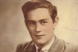
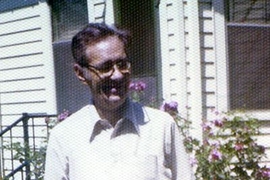
Henry Molaison, the famous amnesic patient better known as “H.M.,” was unable to form new long-term memories following brain surgery to treat his epilepsy. Scientists who studied his condition made groundbreaking discoveries that revealed how memory works, and before his 2008 death, H.M. and his guardian agreed that his brain would be donated to science. One year after his death, H.M.’s brain was sliced into 2,401 70-micron-thick sections for further study.
MIT neuroscience professor emerita Suzanne Corkin studied H.M. during his life and is now part of a team that is analyzing his brain. She is an author of a paper appearing in Nature Communications today reporting preliminary results of the postmortem study. The research team was led by Jacopo Annese at the University of California at San Diego (UCSD).
Q: What can we learn from studying H.M.’s brain after his death? And when did you begin laying the groundwork for these postmortem studies?
A: It was important to get H.M.’s brain after he died, for three reasons: first of all, to document the exact locus and extent of his lesions, in order to identify the neural substrate for declarative memory. Second, to evaluate the status of the intact brain tissue, revealing the possible brain substrates for the many cognitive functions that H.M. performed normally, including nondeclarative learning without awareness. The third reason was to identify any new abnormalities that occurred as a result of his getting old and were unrelated to the operation.
In 1992, I explained to H.M. and his conservator that it would be extremely valuable to have his brain after he died. I told them how important he was to the science of memory, and that he had already made amazing contributions. It would make those even more significant to actually have his brain and see exactly where the damage was. That year, they signed a brain donation form leaving his brain to Massachusetts General Hospital [MGH] and MIT.
In 2002, I assembled a committee to plan what we would do with H.M.’s brain when he died, moment by moment. Detailed planning in advance is necessary if you want to study a brain after death. You can’t wait until the person dies to set this up because it won’t happen. H.M.’s nursing home had a sheet of paper inside his chart telling them exactly what they were supposed to do. Everything went off as planned, and we had him in the scanner at MGH’s Martinos Center for Biomedical Imaging in just under four hours from the time he died. We scanned him for nine hours, and the next morning, MGH neuropathologist Matthew Frosch carried out the autopsy.
A year later, Jacopo Annese’s team at UCSD sliced the brain. I was excited and nervous during this historic event, but the three-day procedure went without a hitch.
Q: What does this new study reveal?
A: This specific report is part of a much broader study that started in 1992 when H.M. signed the brain donation form, and it will continue for years to come. The initial results confirm what we already knew about the size and shape of H.M.’s lesions.
In addition, we found white matter damage related to the original operation, as well as age-related white matter abnormalities. These changes will be elucidated in future histological examinations in which it will be possible to distinguish old white matter damage from new white matter damage.
We discovered a new lesion in the lateral orbital gyrus of the left frontal lobe. This damage was also visible in the postmortem MRI scans. The etiology of this lesion is presently unknown; future histological studies will clarify the cause and timeframe of this damage. Currently, it is unclear whether this lesion had any consequence for H.M.’s behavior.
The Nature Communications paper is a promissory note. It’s the beginning of the second phase of H.M.’s groundbreaking contributions to the science of memory.
Q: What further studies will be done on H.M.’s brain?
A: The first thing that will happen now is that tissue samples will be sent to a group of expert neuropathologists who will conduct a standard neuropathological exam. Those results should be available this year. Another top priority will be the complete neurohistological examination of the entire brain from front to back, top to bottom.
After that, or maybe concurrently with that, individual investigators who have specific research questions to ask about H.M.’s brain will be invited to submit proposals to a committee. If the project seems scientifically worthwhile, then tissue will be sent to those investigators for their examination.
Numerous publications will result over the ensuing years. It’s hard to overestimate how much we will learn from subsequent studies of H.M.’s brain. This study is just whetting people’s appetite. The heavy-duty science will follow.
Preliminary analysis of H.M.’s brain tissue lays groundwork for more comprehensive studies.
Publication Date:
Press Contact:

Caption:
Suzanne Corkin
Credits:
Photo: Patrick Gillooly
Related Topics
Related Articles


