
Faculty Highlight: Ibrahim Cissé
Assistant professor of physics probes the formation of enzyme clusters that enable gene copying and protein production in living cells.
Assistant professor of physics probes the formation of enzyme clusters that enable gene copying and protein production in living cells.
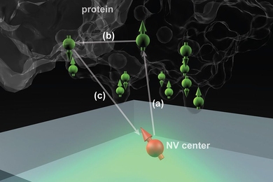
New technique could use tiny diamond defects to reveal unprecedented detail of molecular structures.
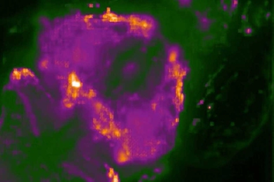
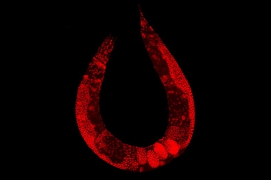
New technique allows scientists to monitor the entire nervous system of a small worm.
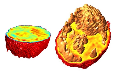
New microscopy technique captures 3-D images of cells as they flow through a microfluidic channel.

Molecular charges affect peptide transport, MIT postdoc Leon Li demonstrates
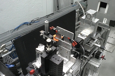
Device could open up new areas of research on materials and biological samples at tiny scales.
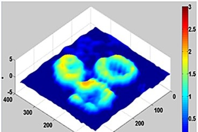
New microscopy technique allows scientists to visualize cells through the walls of silicon microfluidic devices.
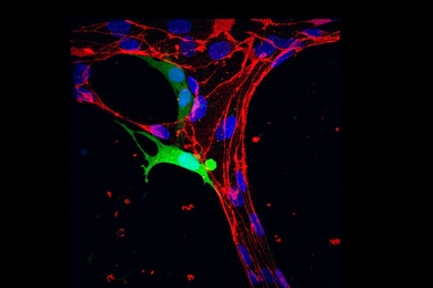
A microfluidic platform provides a high-resolution view of a crucial step in cancer metastasis.
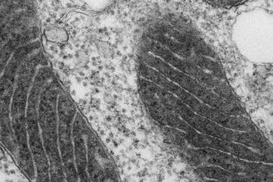
Protein-labeling technique allows high-resolution visualization of molecules inside cells.
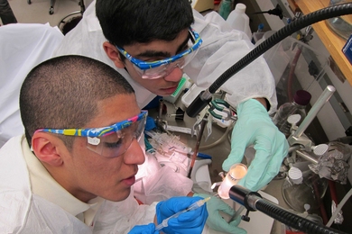
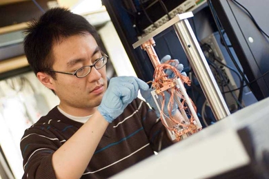
MIT researchers are building a microscope that uses MRI technology to image viruses and other tiny biological structures.
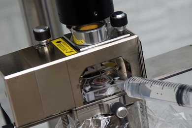
New technique improves researchers’ ability to measure a key property of material surfaces.
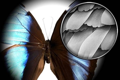
Quantum mechanics could help build ultra-high-resolution electron microscopes that won't destroy living cells, according to MIT electrical engineers.