A newly complete database of human protein kinases and their preferred binding sites provides a powerful new platform to investigate cell signaling pathways.
Culminating 25 years of research, MIT, Harvard University, and Yale University scientists and collaborators have unveiled a comprehensive atlas of human tyrosine kinases — enzymes that regulate a wide variety of cellular activities — and their binding sites.
The addition of tyrosine kinases to a previously published dataset from the same group now completes a free, publicly available atlas of all human kinases and their specific binding sites on proteins, which together orchestrate fundamental cell processes such as growth, cell division, and metabolism.
Now, researchers can use data from mass spectrometry, a common laboratory technique, to identify the kinases involved in normal and dysregulated cell signaling in human tissue, such as during inflammation or cancer progression.
“I am most excited about being able to apply this to individual patients’ tumors and learn about the signaling states of cancer and heterogeneity of that signaling,” says Michael Yaffe, who is the David H. Koch Professor of Science at MIT, the director of the MIT Center for Precision Cancer Medicine, a member of MIT’s Koch Institute for Integrative Cancer Research, and a senior author of the new study. “This could reveal new druggable targets or novel combination therapies.”
The study, published in Nature, is the product of a long-standing collaboration with senior authors Lewis Cantley at Harvard Medical School and Dana-Farber Cancer Institute, Benjamin Turk at Yale School of Medicine, and Jared Johnson at Weill Cornell Medical College.
The paper’s lead authors are Tomer Yaron-Barir at Columbia University Irving Medical Center, and MIT’s Brian Joughin, with contributions from Kontstantin Krismer, Mina Takegami, and Pau Creixell.
Kinase kingdom
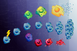
Human cells are governed by a network of diverse protein kinases that alter the properties of other proteins by adding or removing chemical compounds called phosphate groups. Phosphate groups are small but powerful: When attached to proteins, they can turn proteins on or off, or even dramatically change their function. Identifying which of the almost 400 human kinases phosphorylate a specific protein at a particular site on the protein was traditionally a lengthy, laborious process.
Beginning in the mid 1990s, the Cantley laboratory developed a method using a library of small peptides to identify the optimal amino acid sequence — called a motif, similar to a scannable barcode — that a kinase targets on its substrate proteins for the addition of a phosphate group. Over the ensuing years, Yaffe, Turk, and Johnson, all of whom spent time as postdocs in the Cantley lab, made seminal advancements in the technique, increasing its throughput, accuracy, and utility.
Johnson led a massive experimental effort exposing batches of kinases to these peptide libraries and observed which kinases phosphorylated which subsets of peptides. In a corresponding Nature paper published in January 2023, the team mapped more than 300 serine/threonine kinases, the other main type of protein kinase, to their motifs. In the current paper, they complete the human “kinome” by successfully mapping 93 tyrosine kinases to their corresponding motifs.
Next, by creating and using advanced computational tools, Yaron-Barir, Krismer, Joughin, Takegami, and Yaffe tested whether the results were predictive of real proteins, and whether the results might reveal unknown signaling events in normal and cancer cells. By analyzing phosphoproteomic data from mass spectrometry to reveal phosphorylation patterns in cells, their atlas accurately predicted tyrosine kinase activity in previously studied cell signaling pathways.
For example, using recently published phosphoproteomic data of human lung cancer cells treated with two targeted drugs, the atlas identified that treatment with erlotinib, a known inhibitor of the protein EGFR, downregulated sites matching a motif for EGFR. Treatment with afatinib, a known HER2 inhibitor, downregulated sites matching the HER2 motif. Unexpectedly, afatinib treatment also upregulated the motif for the tyrosine kinase MET, a finding that helps explain patient data linking MET activity to afatinib drug resistance.
Actionable results
There are two key ways researchers can use the new atlas. First, for a protein of interest that is being phosphorylated, the atlas can be used to narrow down hundreds of kinases to a short list of candidates likely to be involved. “The predictions that come from using this will still need to be validated experimentally, but it’s a huge step forward in making clear predictions that can be tested,” says Yaffe.
Second, the atlas makes phosphoproteomic data more useful and actionable. In the past, researchers might gather phosphoproteomic data from a tissue sample, but it was difficult to know what that data was saying or how to best use it to guide next steps in research. Now, that data can be used to predict which kinases are upregulated or downregulated and therefore which cellular signaling pathways are active or not.
“We now have a new tool now to interpret those large datasets, a Rosetta Stone for phosphoproteomics,” says Yaffe. “It is going to be particularly helpful for turning this type of disease data into actionable items.”
In the context of cancer, phosophoproteomic data from a patient’s tumor biopsy could be used to help doctors quickly identify which kinases and cell signaling pathways are involved in cancer expansion or drug resistance, then use that knowledge to target those pathways with appropriate drug therapy or combination therapy.
Yaffe’s lab and their colleagues at the National Institutes of Health are now using the atlas to seek out new insights into difficult cancers, including appendiceal cancer and neuroendocrine tumors. While many cancers have been shown to have a strong genetic component, such as the genes BRCA1 and BRCA2 in breast cancer, other cancers are not associated with any known genetic cause. “We’re using this atlas to interrogate these tumors that don’t seem to have a clear genetic driver to see if we can identify kinases that are driving cancer progression,” he says.
Biological insights
In addition to completing the human kinase atlas, the team made two biological discoveries in their recent study. First, they identified three main classes of phosphorylation motifs, or barcodes, for tyrosine kinases. The first class is motifs that map to multiple kinases, suggesting that numerous signaling pathways converge to phosphorylate a protein boasting that motif. The second class is motifs with a one-to-one match between motif and kinase, in which only a specific kinase will activate a protein with that motif. This came as a partial surprise, as tyrosine kinases have been thought to have minimal specificity by some in the field.
The final class includes motifs for which there is no clear match to one of the 78 classical tyrosine kinases. This class includes motifs that match to 15 atypical tyrosine kinases known to also phosphorylate serine or threonine residues. “This means that there’s a subset of kinases that we didn’t recognize that are actually playing an important role,” says Yaffe. It also indicates there may be other mechanisms besides motifs alone that affect how a kinase interacts with a protein.
The team also discovered that tyrosine kinase motifs are tightly conserved between humans and the worm species C. elegans, despite the species being separated by more than 600 million years of evolution. In other words, a worm kinase and its human homologue are phosphorylating essentially the same motif. That sequence preservation suggests that tyrosine kinases are highly critical to signaling pathways in all multicellular organisms, and any small change would be harmful to an organism.
The research was funded by the Charles and Marjorie Holloway Foundation, the MIT Center for Precision Cancer Medicine, the Koch Institute Frontier Research Program via L. Scott Ritterbush, the Leukemia and Lymphoma Society, the National Institutes of Health, Cancer Research UK, the Brain Tumour Charity, and the Koch Institute Support (core) grant from the National Cancer Institute.