Severe traumatic injuries that destroy large volumes of muscle can impact a person’s health, mobility, and quality of life for a lifetime. Promising new research co-led by Ritu Raman, the d’Arbeloff Career Development Assistant Professor of Mechanical Engineering, and MIT collaborators aims to restore mobility for those who have lost muscle through disease or trauma.
“For so many years, I had an idea that if we were able to exercise muscle grafts after they'd been implanted in an injury, we'd be able to keep the graft active [and] prevent it from atrophying by integrating it with the surrounding host tissue,” says Raman.
A new paper published in the journal Biomaterials presents research that shows that targeted actuation of implanted muscle grafts via noninvasive light stimulation restores motor function in mice to levels similar to those of healthy mice by two weeks post-injury.
Volumetric muscle loss, the type of injury this work aims to address, has long been explored in the field of tissue engineering, but many previous studies have focused on creating replacement tissue in the lab, implanting it in the body, and then passively letting the patient’s system integrate the implant. These lab-engineered skeletal muscle implants only offer limited mobility recovery but, in Raman’s study, the mice completely recover functional mobility within two weeks.
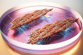
“We engineer optogenetic muscle grafts that contract in response to light and implant these within mice with muscle loss injuries in their hind legs,” explains Raman. “We then ‘exercise’ the implant daily by noninvasively shining light on the mouse's leg through the skin. The approach keeps muscle implants active while they are engrafting with the surrounding host tissue.”
The researchers were excited to discover that actuating grafts seem to turn on cell signals related to the growth of new blood vessels and nerves. This provides a potential explanation for why injured mice are able to recover so completely and so quickly.
“Exercising muscle grafts after they've implanted does more than just make muscle stronger, it also appears to affect how muscle communicates with other tissue, like blood vessels and nerves,” says Raman. “By actively communicating with the implant and exercising the muscle graft, you can actually improve and accelerate recovery timelines.”


