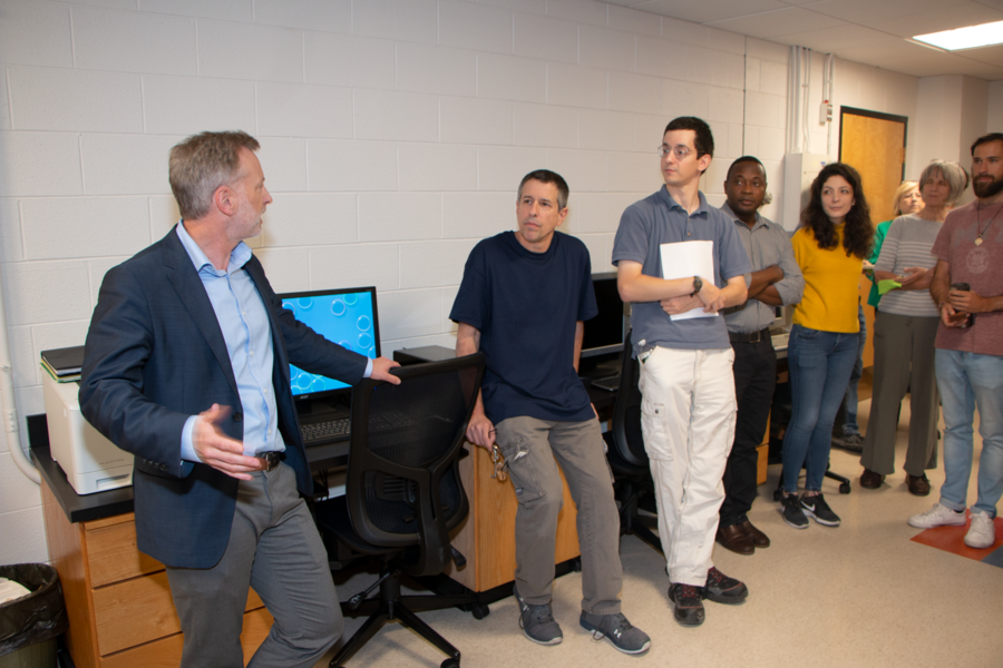
The Materials Research Laboratory (MRL) welcomed about three dozen guests to celebrate renovations to the Electron Microscopy (EM) Shared Experimental Facility in Building 13, the Vannevar Bush Building, last month.
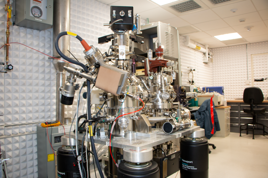
The EM suite, which is part of the National Science Foundation-funded Materials Research Science and Engineering Center (MRSEC) within MRL, is now home to an ultra-high vacuum transmission electron microscope (TEM) and a scanning tunneling microscope (STM), both of which were donated by IBM to MIT.
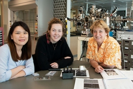
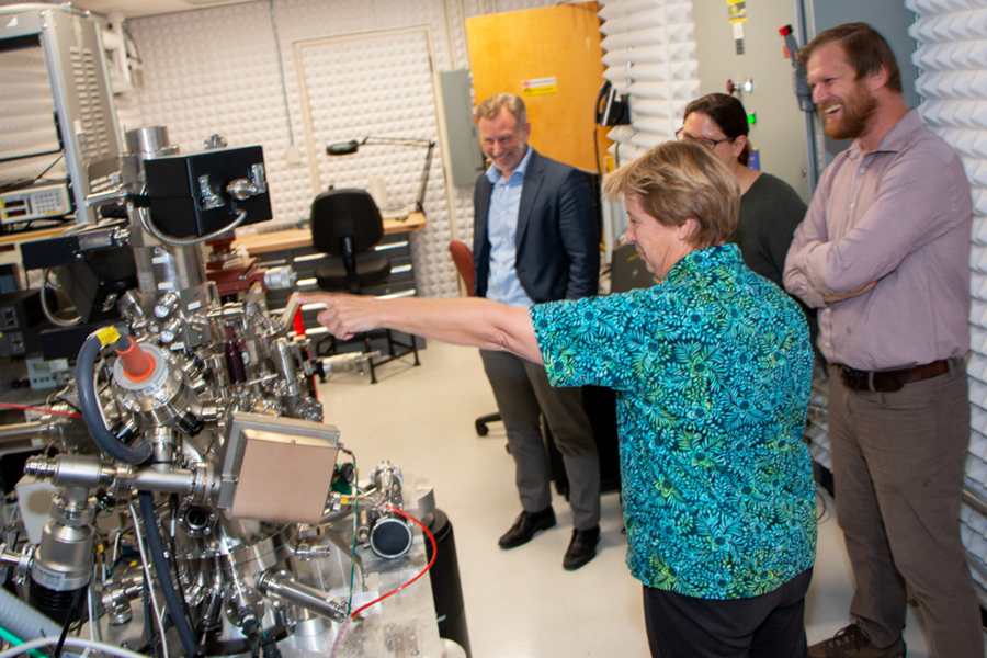
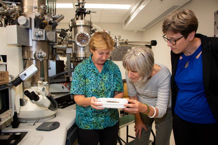
“Part of this welcome party, from my point of view, is to see which of you would like to get involved with this new equipment,” said Frances M. Ross, the Ellen Swallow Richards Professor in Materials Science and Engineering, who joined the Department of Materials Science and Engineering faculty last year, moving from the Nanoscale Materials Analysis Department at the IBM Thomas J. Watson Research Center.
“Both microscopes have unique capabilities. With the TEM, we can grow materials onto a sample while observing the action in the microscope, making a movie that shows how growth takes place. The interior is at ultra-high vacuum so we can prepare a clean sample and be sure that it will stay clean for the duration of the experiment. The STM is also an ultra-high vacuum instrument. It has four tips to measure the sample, capabilities for growth, as well as an integrated ion beam to modify the sample surface.” Ross said her team would be delighted to give tours of the equipment to MIT community members who are interested in learning more.
Looking back on the renovations, Ross says, “You go from a phase where you say, how can this take so long, why must it take months and months and months, and then at other times, you say, wow, how did they possibly finish it so quickly?”
Greene Construction completed the EM suite renovations, which included new flooring and lighting, a new entrance, repainting, and an updated meeting area with video presentation capability for meetings or teaching.
“It’s a very exciting time for electron microscopy at MIT,” MRL Co-Director Geoffrey S.D. Beach says. “The opportunity is not just to renew this facility, but, as you know, Frances Ross has brought some remarkably unique equipment from IBM and embedded that within this shared experimental facility … We expect great things to happen.”
The electron microscopy suite is one of several MRSEC Shared Experimental Facilities, which also include materials analysis (surface, thermal, and optical), X-ray diffraction, and nanostructured materials.