
The stiffness or elasticity of a cell can reveal much about whether the cell is healthy or diseased. Cancer cells, for instance, are known to be softer than normal, while asthma-affected cells can be rather stiff.
Determining the mechanical properties of cells may thus help doctors diagnose and track the progression of certain diseases. Current methods for doing this involve directly probing cells with expensive instruments, such as atomic force microscopes and optical tweezers, which make direct, invasive contact with the cells.
Now MIT engineers have devised a way to assess a cell’s mechanical properties simply by observation. The researchers use standard confocal microscopy to zero in on the constant, jiggling motions of a cell’s particles — telltale movements that can be used to decipher a cell’s stiffness. Unlike optical tweezers, the team’s technique is noninvasive, running little risk of altering or damaging a cell while probing its contents.
“There are several diseases, like certain types of cancer and asthma, where stiffness of the cell is known to be linked to the phenotype of the disease,” says Ming Guo, the Brit and Alex d'Arbeloff Career Development Assistant Professor in MIT’s Department of Mechanical Engineering. “This technique really opens a door so that a medical doctor or biologist, if they would like to know the material property of cell in a very quick, noninvasive way, can now do it.”
Guo and graduate student Satish Kumar Gupta have published their results in the Journal of the Mechanics and Physics of Solids.
Stirring spoons
In his 1905 PhD thesis, Albert Einstein derived a formula, known as the Stokes-Einstein equation, that makes it possible to calculate a material’s mechanical properties by observing and measuring the movement of particles in that material. There’s just one catch: The material must be “in equilibrium,” meaning that any particle motions must be due to the effect of the material’s temperature rather than any external forces acting on the particles.
“You can think of equilibrium as being a hot cup of coffee,” Guo says. “The coffee’s temperature alone can drive sugar to disperse. Now if you stir the coffee with a spoon, the sugar dissolves faster, but the system is not driven solely by temperature any more and is no longer in equilibrium. You’re changing the environment, putting energy in and making the reaction happen faster.”
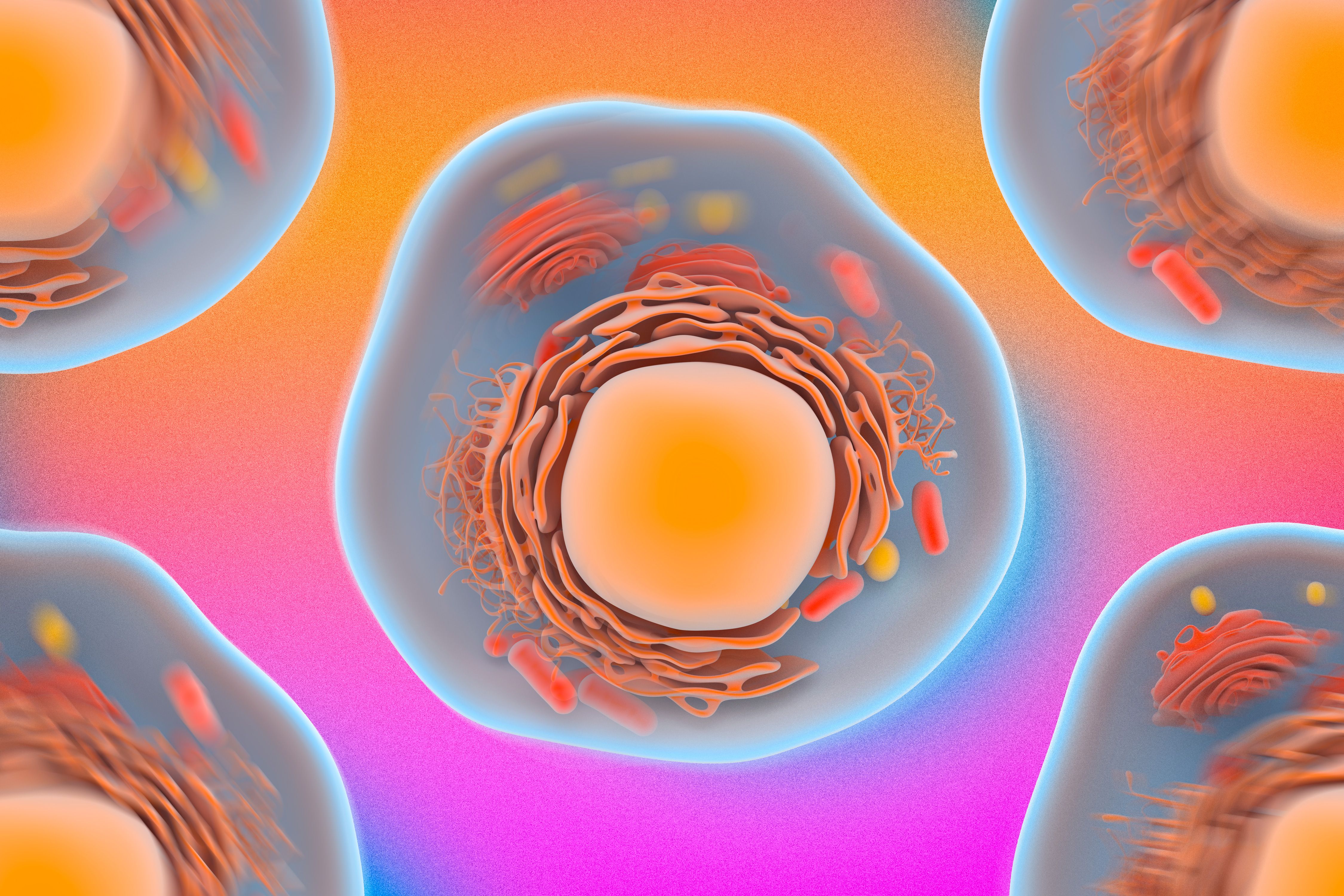
Within a cell, organelles such as mitochondria and lysosomes are constantly jiggling in response to the cell’s temperature. However, Guo says, there are also “many minispoons” stirring up the surrounding cytoplasm, in the form of proteins and molecules that, every so often, actively push vibrating organelles around like billiard balls.
The constant blur of activity in a cell has made it difficult for scientists to discern, simply by looking, which motions are due to temperature and which are due to more active, “spoon-like” processes. This limitation, Guo says, has “basically shut the door on using Einstein’s equation and pure observation to measure a cell’s mechanical properties.”
Frame by frame
Guo and Gupta surmised that there might be a way to tease out temperature-driven motions in a cell by looking at the cell within a very narrow timeframe. They realized that particles energized solely by temperature exhibit a constant jiggling motion. No matter when you look at a temperature-driven particle, it’s bound to be moving.
In contrast, active processes that can knock a particle around a cell’s cytoplasm do so only occasionally. Seeing such active movements, they hypothesized, would require looking at a cell over a longer timeframe.
To test their hypothesis, the researchers carried out experiments on human melanoma cells, a line of cancer cells they chose for their ability to grow easily and quickly. They injected small polymer particles into each cell, then tracked their motions under a standard confocal fluorescent microscope. They also varied the cells’ stiffness by introducing salt into the cell solution — a process that draws water out of cells, making them more compressed and stiff.
The researchers recorded videos of the cells at different frame rates and observed how the particles’ motions changed with cell stiffness. When they watched the cells at frequencies higher than 10 frames per second, they mostly observed particles jiggling in place; these vibrations appeared to be caused by temperature alone. Only at slower frame rates did they spot more active, random movements, with particles shooting across wider distances within the cytoplasm.
For each video, they tracked the path of a particle and applied an algorithm they had developed to calculate the particle’s average travel distance. They then plugged this motion value into a generalized format of the Stokes-Einstein equation.
Guo and Gupta compared their calculations of stiffness with actual measurements they made using optical tweezers. Their calculations matched up with measurements only when they used the motion of particles captured at frequencies of 10 frames per second and higher. Guo says this suggests that particle motions occurring at high frequencies are indeed temperature-driven.
The team’s results suggest that if researchers observe cells at fast enough frame rates, they can isolate particle motions that are purely driven by temperature, and determine their average displacement — a value that can be directly plugged into Einstein’s equation to calculate a cell’s stiffness.
“Now if people want to measure the mechanical properties of cells, they can just watch them,” Guo says.
The team is now working with doctors at Massachusetts General Hospital, who hope to use the new, noninvasive technique to study cells involved in cancer, asthma, and other conditions in which cell properties change as a disease progresses.
“People have an idea that structure changes, but doctors want to use this method to demonstrate whether there is a change, and whether we can use this to diagnose these conditions,” Guo says.
This research was funded, in part, by MIT’s Department of Mechanical Engineering.