
For many decades, neuroscientists believed there was a “critical period” in which the brain could learn to make sense of visual input, and that this window closed around the age of 6 or 7.
Recent work from MIT Professor Pawan Sinha has shown that the picture is more nuanced than that. In many studies of children in India who had surgery to remove congenital cataracts beyond the age of 7, he has found that older children can learn visual tasks such as recognizing faces, distinguishing objects from a background, and discerning motion.
In a new study, Sinha and his colleagues have now discovered anatomical changes that occur in the brains of these patients after their sight is restored. These changes, seen in the structure and organization of the brain’s white matter, appear to underlie some of the visual improvements that the researchers also observed in these patients.
The findings further support the idea that the window of brain plasticity, for at least some visual tasks, extends much further than previously thought.
“Given the remarkable level of remodeling of brain structure that we are seeing, it reinforces the point that we have been trying to make with our behavioral results, that all children ought to be provided treatment,” says Pawan Sinha, an MIT professor of brain and cognitive sciences and one of the authors of the study.
Bas Rokers, an associate professor and director of the Neuroimaging Center at New York University Abu Dhabi, is the senior author of the study, which appears this week in the Proceedings of the National Academy of Sciences. The paper’s lead authors are Caterina Pedersini, a postdoc at New York University Abu Dhabi; Nathaniel Miller, who is studying medicine at the University of Minnesota Medical School; and Tapan Gandhi, a former postdoc in the Sinha Lab who is now an associate professor at the Indian Institute of Technology. Sharon Gilad-Gutnick, an MIT research scientist, and Vidur Mahajan, director of the Center for Advanced Research on Imaging, Neuroscience, and Genomics, are also authors of the paper.
White matter plasticity
In developed nations such as the United States, infants born with cataracts are treated within a few weeks of birth. However, in developing nations such as India, a higher percentage of these cases go untreated.
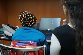
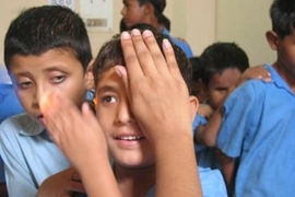
Nearly 20 years ago, Sinha launched an initiative called Project Prakash, with the mission to offer medical treatment to blind and vision-impaired children in India. Each year, the project screens thousands of children, many of whom are provided with glasses or more advanced interventions such as surgical removal of cataracts. Some of these children, with their families’ permission, also participate in studies of how the brain’s visual system responds after sight is restored.
In the new study, the researchers wanted to explore whether they could detect any anatomical changes in the brain that might correlate with the behavioral changes that they have previously seen in children who received treatment. They scanned 19 participants, ranging in age from 7 to 17 years of age, at several time points after they had surgery to remove congenital cataracts.
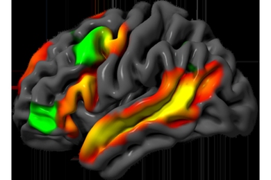
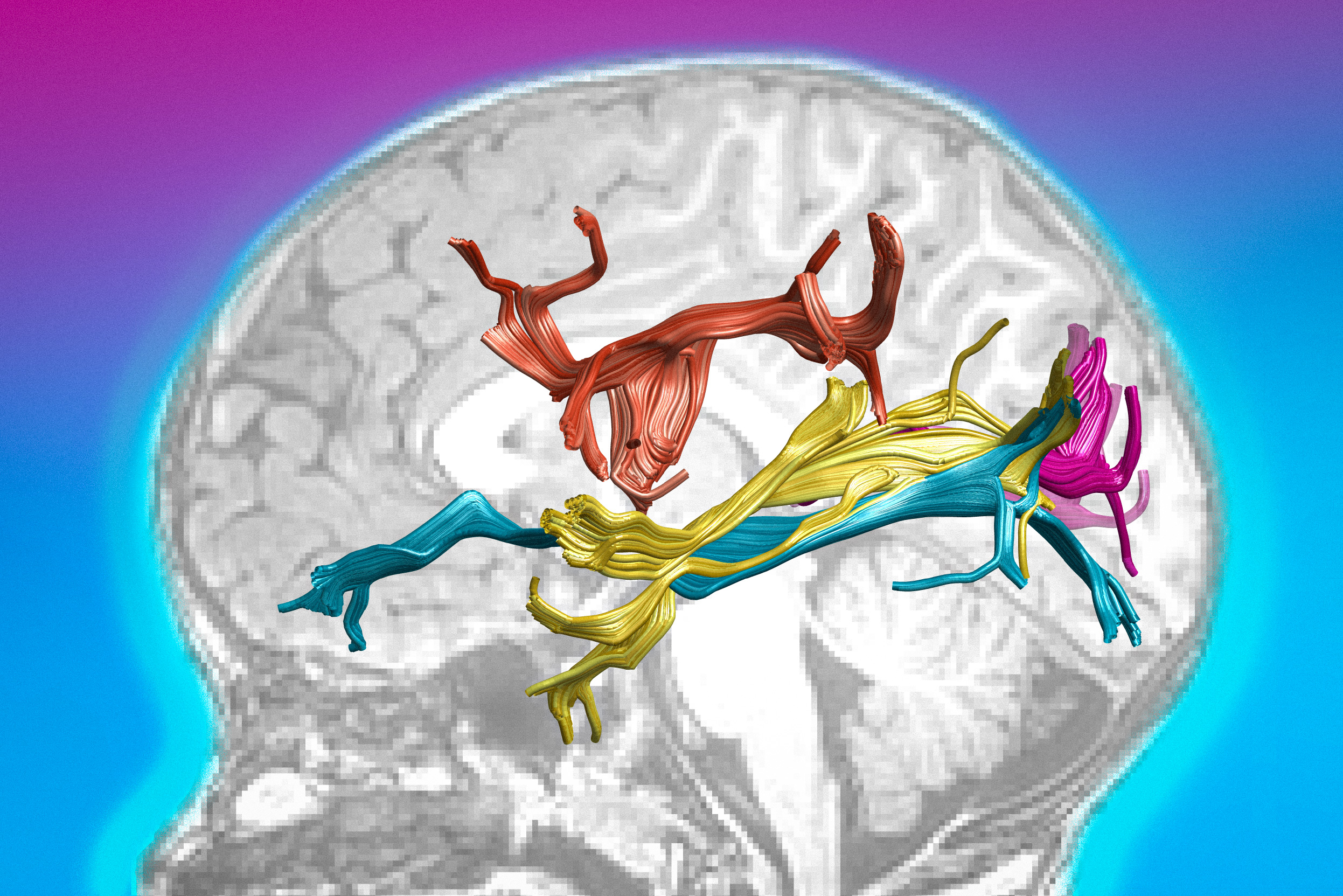
To analyze anatomical changes in the brain, the researchers used a specialized type of magnetic resonance imaging called diffusion tensor imaging. This type of imaging can reveal changes in the organization of the white matter — bundles of nerve fibers that connect different regions of the brain.
Diffusion tensor imaging, which tracks the movement of hydrogen nuclei in water molecules, produces two measurements: mean diffusivity, a measure of how freely water molecules can move, and fractional anisotropy, which reveals the extent to which water is forced to move in one direction over another.
An increase in fractional anisotropy suggests that water molecules are more constrained because nerve fibers in the white matter are oriented in a particular direction.
“If you see increasing fractional anisotropy and decreasing mean diffusivity, then you can infer that what’s happening is that the nerve fibers are growing in volume and they’re getting more organized in terms of their alignment,” Sinha says. “When we look at the white matter of the brain, then we see precisely these kinds of changes in some of the white matter bundles.”
The researchers observed these changes specifically in white matter pathways that are part of the later stages of the visual system, which is believed to be involved in higher-order functions such as face perception. These improvements occurred gradually over several months following the surgery.
“You see anatomical changes in the white matter, but in separate studies using functional neuroimaging, you also see increasing specialization, as a function of visual experience, similar to what happens in typical development,” Gilad-Gutnick says.
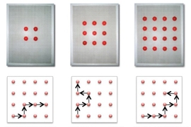
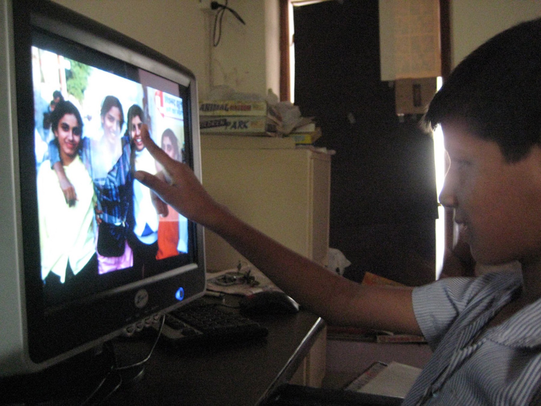
The researchers also tested the participants’ performance on a variety of visual tasks and found that their ability to distinguish faces from other objects was correlated with the amount of structural change in the white matter pathways associated with higher-order visual function.
In comparison, while the treated children showed some improvements in visual acuity — the ability to clearly see details of objects at a distance — their acuity never fully recovered, and they showed only minimal changes in the white matter organization of the early visual pathways.
“The notion that plasticity is a time-limited resource and that past a certain window we can’t expect much improvement, that does seem to hold true for low-level visual function like acuity,” Sinha says. “But when we talk about a higher-order visual skill, like telling a face from a non-face, there we do see behavioral improvements over time, and we also find there to be a correlation between the improvement that we are seeing behaviorally and the changes that we see anatomically.”
Benefits of treatment
The researchers also found that children who had cataracts removed at a younger age showed greater, and faster, gains in face-perception ability than older children. However, all of the children showed at least some improvement in this skill, along with changes in the structure of the white matter.
The findings suggest that older children can benefit from this kind of surgery and offers further evidence that it should be offered to them, Sinha says.
“If the brain has such outstanding abilities to reconfigure itself and even to change its structure, then we really ought to capitalize on that plasticity and provide children with treatment, irrespective of age,” he says.
Sinha’s lab is now analyzing additional imaging data from Project Prakash patients. In one study, the researchers are investigating whether the patients show any changes in the thickness of their gray matter, especially in the brain’s sensory processing areas, after treatment. The researchers are also using functional MRI to try to localize visual functions such as face perception, to see if they arise in the same parts of the brain that they do in people born with normal sight.
The research was funded by the National Eye Institute.