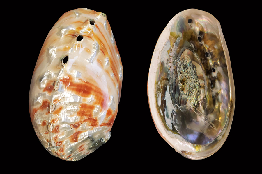
Nacre, the iridescent material that lines mollusk shells such as mother-of-pearl and abalone, has long been a prized find of beachcombers and shell collectors, due to the natural beauty and variety of color that can be found therein. But scientists and engineers have also long marveled at and studied nacre; it’s a tough and strong material, composed of alternating layers of aragonite platelets and organic protein-based film. The natural world contains many materials that have evolved over time to optimize strength, durability, and performance. As researchers and engineers look to develop improved and more sustainable building materials, they are increasingly looking to nature for inspiration.
The physical makeup of nacre allows it to withstand considerable amounts of pressure and damage along the platelets without causing major damage throughout the whole shell. It has been supposed by some that more is at play of the individual platelets that allows them such extraordinary strength and durability, but researchers have lacked the tools and processes to dig deeper into the relationship between the crystal orientation and the mechanical properties — until now.
Over the past two decades, the shells have typically been tested for their strength using techniques such as macroscopic bending test, micro-/nano-indentation, and atomic force microscope. Now, MIT assistant professor of civil and environmental engineering Admir Masic, graduate student Hyun-Chae “Chad” Loh, and five others have combined scanning electron microscopy and micro-indentation with Raman spectroscopy and developed a powerful chemo-mechanical characterization method that allows three-dimensional stress and strain mapping through a technique known as piezo-Raman.
“We developed a methodology to extract important chemo-mechanical information from a biological system that is very well known and studied,” explains Masic, whose findings were recently published in Communications Materials. “Correlating micro-indentation and piezo-Raman results allowed us to evaluate and quantify the amount of stress dissipated through the hierarchical structure.”
The new approach to quantifying the mechanical performance of the material is enough to be big news on its own, but during the process, Masic and fellow researchers — whom he credits with much of the work in this collaborative effort — were surprised by the results.
“We first applied these tools to study the strain-hardening mechanism at a few microns scale. However, we noticed that the dissipation of energy was not confined to the brick-and-mortar structure, but was affecting a much larger area than we expected. We expanded our scope of study to a larger scale and found this new toughening mechanism that is related to a mesostructure on a scale of 20 microns,” says Loh. What the researchers found is that stacks of co-oriented aragonite platelets constitute another hierarchical level of structure, which toughens the material as it is stressed.
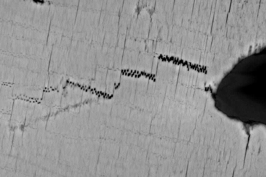
Polarized Raman, another technique used in this study, helped the team observe what’s known as the crystallographic orientation of the aragonite bricks. Through the investigation of the orientation patterns, researchers were able to elucidate the characteristic length scale of the aragonite stacks and relate it to the crack propagation patterns. The cracks propagated between the aragonite stacks, evincing their mechanical contribution to nacre’s toughness.
“This gave us an opening for potentially explaining what is causing this toughening at the larger scales. Systematic arrangements of crystals can be found within other biomineral materials, such as our teeth, and the micro-texture of the materials directly impacts their function.” says Masic.
Mimicking natural materials like nacre has been a popular strategy for designing new materials. The small scale of their structures, however, poses a challenge for replicating and manufacturing the natural morphologies. “With our discovery, we propose a new biomimicry strategy of simulating nacre’s structure on a 10-micron or bigger scale, instead of the nano level.” says Masic.
It's exciting news for researchers who are exploring new possibilities for synthetic materials inspired by natural design.
This research was funded, in part, by Kwanjeong Educational Foundation.