Overcoming limitations of super-resolution microscopy to optimize imaging of RNA in living cells is a key motivation for physics graduate student Takuma Inoue, who works in the lab of MIT assistant professor of physics Ibrahim Cissé.
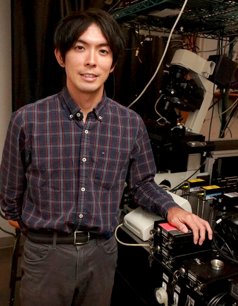
Inoue, 26, was the first student to join Cissé's lab at MIT in January 2014, and he built the lab's super-resolution microscopy setup to study enzyme clusters that enable gene copying and protein production within living cells. Inoue, who this September enters his fourth year toward his PhD, originally started his experimental work in an atomic physics lab, where he worked on an imaging setup to trap extremely cold atoms in a vacuum. He is studying biophysics, atomic physics, and condensed matter physics.
After learning that Cissé needed someone to set up his super-resolution microscopy, Inoue switched to Cissé's lab. Because he did not have a biology background, Inoue says, "I wasn't very much familiar with that, but the tools that you use and the methods for imaging are very common with what I had previously done. By building the setup, I got used to what things we can do in the lab. Then I made the transition to actually targeting some biomolecules within the cell to image and for me that was RNA."
"Initially, I had to start with building the microscope, which took me several months, and then I tried doing the continuation of his previous work which is imaging some kind of protein inside of a living cell," Inoue says. "But then he gave (me) this project of RNA imaging as my main project for my PhD, because that will be more challenging, and we thought no one has ever achieved this big goal."
"My project makes sure that we overcome as many limitations as possible because there are different aspects of this for all the projects that we do," Inoue adds. "There is the setup and there is this data analysis software and also we need to label the target molecules of interest properly. Each of them is, of course, not perfect. There are many challenges and limitations. But if you have a final goal in your project, I think you need to care about all of those different aspects and try optimizing. I've been doing experiments for a long time, so overcoming such limitations in the lab was one of my interests."
Inoue is developing techniques for easily tagging and visualizing RNA directly in living cells. "For me as an experimentalist, it's a very exciting challenge to achieve the imaging of RNA within a live cell and to bring it to the level of a single molecule. My goal is to achieve a technique to image single molecules of RNA inside of a living cell. That can have very broad applications. I think it's very transformative," Inoue says.
The common approach to such imaging is genetic modification that adds a derivative of green fluorescent protein to the target of study — for example, RNA polymerase II. Inoue says his approach is to avoid genetic modification by developing oligonucleotide probes, which are short strands of genetic material that can bind to the target. "I try to deliver these probes into these natural cells and try to see if the target molecules get this fluorescence. And then I bring those cells to the imaging room and then do imaging," he says. The technique is called fluorescent in situ hybridization. The oligonucleotide and the RNA target both start out as single strand molecules, but when they bind they can form a double helix like DNA, Inoue explains.
"There already are approaches for looking at RNA inside dead cells. That's I think the easy part," Takuma's mentor, Cissé, explains. "A handful of labs have also reported on promising ways of labeling RNA in living cells, but those require extensive genetic modifications. Takuma's whole point is actually bringing new techniques for easily tagging and visualizing any arbitrary RNA, without genetic modification, and directly inside the living cell. And his preliminary demonstrations also, I think, look very promising."
Inoue has high hopes for the project. "This project is about labeling arbitrary RNA that exist inside a living cell, and I am at the developing stage of these techniques," he says. "I'm hoping that through this project I can contribute and help many researchers in studying their RNAs of interest and also I, myself, am interested in studying different kinds of RNA."
The Cissé lab's single molecule studies of the role that enzymes, proteins, and RNA play in gene expression is funded under National Institutes of Health Project No. 1DP2CA195769-01 with additional funds from the National Cancer Institute.
The super-resolution imaging setup captures images through the microscope onto an electron-multiplying charge-coupled device (EM-CCD). "It can detect very sensitive signals, even single photons, and also it's a very fast camera," Inoue explains. The EM-CCD has millisecond exposure times but overall it takes several minutes to get one super-resolution image made from about 10,000 images.
A native of Yokohama, Japan, Inoue moved to the U.S. at age 18, three years after his father, Hiroshi Inoue, accepted a position in Maryland starting up a life sciences subsidiary for Canon. Now a resident of Rockville, Maryland, Takuma Inoue received his bachelor's in physics with a minor in mathematics at the University of Maryland at College Park, where his father currently holds the title of Professor of the Practice in Bioengineering.
"I've got a lot of influence from my dad because he was initially an engineer, but then it was a very big surprise to me that he switched to biology and started doing some kind of engineering that could help biologists or could help people. So, that was maybe one of the key events in my life," he says.
"I like to think about scientific challenges and also there are many engineering challenges, and I really like that I am doing that, and also that I am trying to solve the world's most interesting problems in the field of biology using my physics background. I want to see first how this project goes, and if possible, I'd like to continue doing research, and I hope that my career becomes exciting," Inoue says.