Magnetic resonance imaging (MRI) devices can scan the inside of the body in intricate detail, allowing clinicians to spot even the earliest signs of cancer or other abnormalities. But they can be a long and uncomfortable experience for patients, requiring them to lie still in the machine for up to 45 minutes.
Now this scan time could be cut to just 15 minutes, thanks to an algorithm developed at MIT’s Research Laboratory of Electronics.
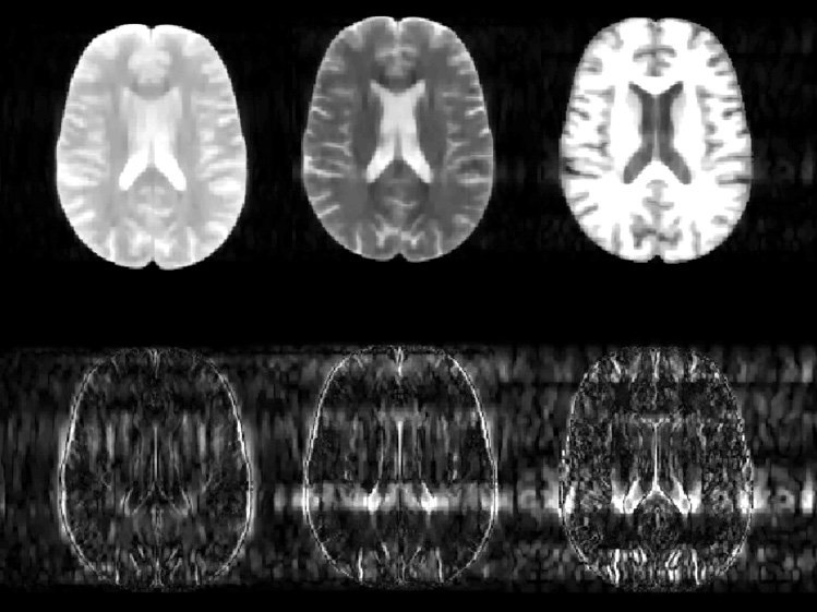
MRI scanners use strong magnetic fields and radio waves to produce images of the body. Rather than taking just one scan of a patient, the machines typically acquire a variety of images of the same body part, each designed to create a contrast between different types of tissue. By comparing multiple images of the same region, and studying how the contrasts vary across the different tissue types, radiologists can detect subtle abnormalities such as a developing tumor. But taking multiple scans of the same region in this way is time-consuming, meaning patients must spend long periods inside the machine.
In a paper to be published in the journal Magnetic Resonance in Medicine, researchers led by Elfar Adalsteinsson, an associate professor of electrical engineering and computer science and health sciences and technology, and Vivek Goyal, the Esther and Harold E. Edgerton Career Development Associate Professor of Electrical Engineering and Computer Science, detail an algorithm they have developed to dramatically speed up this process. The algorithm uses information gained from the first contrast scan to help it produce the subsequent images. In this way, the scanner does not have to start from scratch each time it produces a different image from the raw data, but already has a basic outline to work from, considerably shortening the time it takes to acquire each later scan.
To create this outline, the software looks for features that are common to all the different scans, such as the basic anatomical structure, Adalsteinsson says. “If the machine is taking a scan of your brain, your head won’t move from one image to the next,” he says. “So if scan number two already knows where your head is, then it won’t take as long to produce the image as when the data had to be acquired from scratch for the first scan.”
In particular, the algorithm uses the first scan to predict the likely position of the boundaries between different types of tissue in the subsequent contrast scans. “Given the data from one contrast, it gives you a certain likelihood that a particular edge, say the periphery of the brain or the edges that confine different compartments inside the brain, will be in the same place,” Adalsteinsson says.
However, the algorithm cannot impose too much information from the first scan onto the subsequent ones, Goyal says, as this would risk losing the unique tissue features revealed by the different contrasts. “You don’t want to presuppose too much,” he says. “So you don’t assume, for example, that the bright-and-dark pattern from one image will be replicated in the next image, because in fact those kinds of dark and light patterns are often reversed, and can reveal completely different tissue properties.”
So for each pixel, the algorithm calculates what new information it needs to construct the image, and what information — such as the edges of different types of tissue — it can take from the previous scans, says graduate student and first author Berkin Bilgic.
The result is an MRI scan that is three times quicker to complete, cutting the time patients spend in the machine from 45 to 15 minutes. This faster scan time does have a slight impact on image quality, Bilgic admits, but it is much better than competing algorithms.
The team is now working to further improve the algorithm by speeding up the time it takes to process the raw image data into a final scan that can be analyzed by clinicians, once the patient has stepped out of the MRI machine. Using standard computer processors, this final step currently takes considerably longer than with conventional MRI scans.
But the researchers believe they can reduce this calculation time down to the same as that of conventional MRI scans using recent advances in computing hardware from the gaming industry. “Graphics processing units, or GPUs, are orders of magnitude faster at certain computational tasks than general processors, like the particular computational task that we need for this algorithm,” Adalsteinsson says.
A student at the laboratory is now working to implement the algorithm on a dedicated GPU, he says.
Dwight Nishimura, the director of the Magnetic Resonance Systems Research Laboratory at Stanford University, says Adalsteinsson's group has done some very interesting algorithmic work. “This work is potentially of high significance because it applies to routine clinical MRI, among other applications,” he says. “Ultimately, their approach might enable a substantial reduction in examination time.”
Faster scans could reduce the time patients spend in the machine from 45 to 15 minutes.
Publication Date:
Press Contact:
Credits:
Image credit: courtesy of the researchers