MIT bioengineers have devised a new technique that makes it possible to learn more about how cells are organized in tissues and potentially even to regrow cells for repairing areas of the body damaged by disease, accidents or aging.
The method gives them unprecedented control over organizing cells outside the body in three dimensions, which is how they exist inside the body. It uses electricity to move cells into a desired position, followed by light to lock them into place within a gel that resembles living tissue.
Cells traditionally have been studied in two dimensions in a Petri dish, but certain cells behave differently in two dimensions than in three.
"We have shown that the behavior of cartilage cells is affected significantly when they are organized in 3-D," as is the behavior of other types of cells like stem cells, said MIT Associate Professor Sangeeta Bhatia of the Harvard-MIT Division of Health Sciences and Technology (HST), one author of a paper on the technique due to appear in the May issue of Nature Methods.
"This raises questions about how cells might sense their organization in 3-D and how important this might be in other tissues," said Dirk Albrecht, a postdoctoral associate in Bhatia's lab and lead author of the paper. "We now have a method to answer some of these questions in the lab."
Scientists have until now studied cells in 3-D by placing them randomly into a gel. The cells clump together into "cell spheroids," but that is a slow process, and the size and shape of the cell clumps vary significantly. In addition, cells that communicate by direct contact can end up too far apart.
The new technique allows for precise control of cell organization, and takes minutes to perform compared to hours or days for the other method.
Albrecht and his colleagues have been using a micropatterning technique to carefully position the cells within about 10 microns of each other. That's nearly the diameter of a cell and about one-fifth the diameter of a human hair. The technique uses a device made with photolithography, the same process used to create circuit patterns on electronic microchips.
In the paper, the MIT researchers said they have formed more than 20,000 cell clusters with precise sizes and shapes within a single gel. They have since scaled that up several-fold. They also have created layers of different cells, attempting to mimic the structure of tissue inside the body.
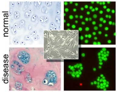
While the technique may one day be applied to engineer tissues for medical applications, its first use will be for basic research on how cells are organized, how they function and communicate in tissues, and how they develop into organs or tumors. The 3-D organization of cells also may help researchers understand how cells respond to drugs when they are in a normal state compared to a diseased state like cancer.
"We also think this technique will be useful for building engineered tissues in specific ways," Bhatia said. "It wasn't possible until now to get this degree of control over cells in 3-D."
Other authors on the paper are MIT HST postdoctoral fellow Greg Underhill, University of California at San Diego Professor of Bioengineering Robert Sah and UCSD alumnus Travis Wassermann.
The authors have applied for a patent on their work.
The research was funded by The Whitaker Foundation, the National Science Foundation, the National Institutes of Health, the David and Lucille Packard Foundation and NASA.
A version of this article appeared in MIT Tech Talk on April 26, 2006 (download PDF).