
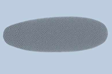
A “scientific sandbox” lets researchers explore the evolution of vision systems
The AI-powered tool could inform the design of better sensors and cameras for robots or autonomous vehicles.
The AI-powered tool could inform the design of better sensors and cameras for robots or autonomous vehicles.
The approach could apply to more complex tissues and organs, helping researchers to identify early signs of disease.

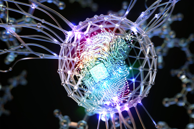
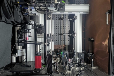
The DIGIT imaging tool could enable the design of quantum devices and shed light on atomic-scale processes in cells and tissues.

A proposed telescope made of thousands of tiny, identical satellites will work to reveal low-frequency radio waves in space.
Acting as a “virtual spectrometer,” SpectroGen generates spectroscopic data in any modality, such as X-ray or infrared, to quickly assess a material’s quality.
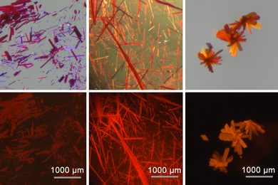
The new dyes are based on boron-containing molecules that were previously too unstable for practical use.

By enabling rapid annotation of areas of interest in medical images, the tool can help scientists study new treatments or map disease progression.

The method enhances 3D bioprinting capabilities, accelerating process optimization for real-world applications in tissue engineering.

MIT CSAIL researchers developed a tool that can model the shape and movements of fetuses in 3D, potentially assisting doctors in finding abnormalities and making diagnoses.

Inventions that protect US service members, advance computing, and enhance communications are recognized among the year's most significant new products.
An international collaboration of neuroscientists, including MIT Professor Ila Fiete, developed a brain-wide map of decision-making at cellular resolution in mice.

By directly imaging material failure in 3D, this real-time technique could help scientists improve reactor safety and longevity.
By combining several cutting-edge imaging technologies, a new microscope system could enable unprecedentedly deep and precise visualization of metabolic and neuronal activity, potentially even in humans.

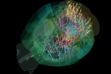
Researchers developed a tool to recreate cells’ family trees. Comparing cells’ lineages and locations within a tumor provided insights into factors shaping tumor growth.
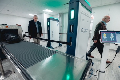
Lincoln Laboratory's 3D microwave imaging technology for detecting concealed threats was integrated into HEXWAVE, commercially developed by Liberty Defense.