Many of us have gotten an X-ray at one time or another, either at the dentist’s or the doctor’s office. Now, astronauts orbiting Earth have shown it’s possible to take an X-ray in space. The effort will help future space explorers diagnose and monitor medical conditions, from fractures and sprains to signs of bone decalcification, while in orbit.
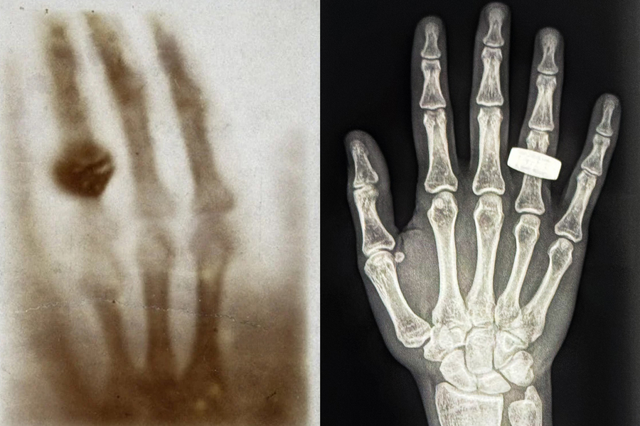
Last week, crew members aboard the Fram2 mission posted to social media and shared the first-ever medical X-ray image taken in space. The image is a black-and-white scan of a hand with a ring, echoing the very first X-ray image ever taken, 130 years ago, by the physicist Wilhelm Roentgen, of his wife’s hand. The new X-ray image was taken in microgravity, inside a four-person space capsule flying at orbital speeds of 17,500 miles per hour, about 200 miles above the Earth’s surface.
The in-flight body scan was part of the SpaceXray project, one of 22 science experiments that astronauts conducted during the Fram2 mission. Operated by SpaceX, Fram2 was the first human spaceflight mission to travel in a polar orbit, looping around the planet from pole to pole. Fram2 gets its mission name from the Norwegian ship “Fram,” which was the first to carry explorers to the Arctic and Antarctic regions in the late 19th century.
The body scans are a first demonstration that medical X-ray imaging can be done within the confines and conditions in space. Lonnie Petersen, a co-investigator on the SpaceXray project, is an associate professor in MIT’s Department of Aeronautics and Astronautics who studies space physiology and the effects of spaceflight on the human body. Petersen helped to define and design the protocol around the SpaceXray project, in collaboration with institutional partners such as Stanford University and the Mayo Clinic, and X-ray hardware companies KA and MinXray. Petersen talked with MIT News about how these first in-orbit X-ray images can help enable safe and healthy longer-term missions in space.
Q: What are the challenges in taking an X-ray in space, versus here on Earth?
A: There are several challenges regarding the hardware, methods, and subjects being X-rayed.
To get hardware certified for spaceflight, it should be miniaturized and as lightweight as possible. There are also increased safety requirements because all devices work in a confined space. The increased requirements drive technology development. I always say space is our best technology accelerator — this is also true for medical technology.
For this project we used a portable, specialized X-ray generator and detector developed by MinXray and KA Imaging for the battlefield and made it applicable for spaceflight.
In terms of methods, one of my concerns was that the increased background radiation might reduce the quality of the image so that it would fall below clinical standards. From the first images we have received from space, it seems that the quality is great. I am very excited to further analyze the full set of images.
We want the X-rays to travel straight through the body part of interest. This requires alignment of equipment and patient. As you can imagine, a floating subject will be harder to position. We will be quantifying any potential impact of this and using it for future updates.
We also do not have radiologists or technicians in space. The methods need to be simple and robust enough for a layperson to operate them.
And, finally, regarding subjects: Entry into space has huge impact on the human body. Blood and fluid are no longer pulled down toward the feet by gravity; they are evenly distributed, and thus there are regional pressures and perfusion changes. The cardiovascular system and the brain are impacted by this over time. Mechanical unloading of the body leads to muscle atrophy and bone decalcification as well reduction in exercise capacity. This mission was only 3.5 days, so the crew will likely not have experienced many negative effects, but with an X-ray, we can now monitor bone health in space. We have never been able to do that before. We can monitor potential fluid buildup in the lungs or check for diseases in the abdomen.
I’ll also take off my physician hat and put on my engineering hat: X-rays are a useful tool in nondestructive hardware tests in aviation (and other areas). This project increases our diagnostic capabilities in space, not just for patients, but also for hardware.
Q: How did the Fram2 crew do it?
A: The crew learned how to take X-rays in one afternoon. It was done as a train-the-trainer model. The protocol was created in advance and the crew took images of each other, checked the quality, and stored the images. We have only seen one image so far, but from that, we are very impressed with the quality, the skills, and the dedication to advancing science by the crew.
Q: What will you learn from these first images?
A: First and foremost, this was a technology demonstration: Can we even do this in space? We are looking forward to analyzing all the images, but from preliminary data it looks like we absolutely can. Now comes a detailed analysis to tease out all the lessons we possibly can learn from this both with regard to current capabilities but also the next steps. The team is, of course, very excited to carry this research forward and break even more ground.