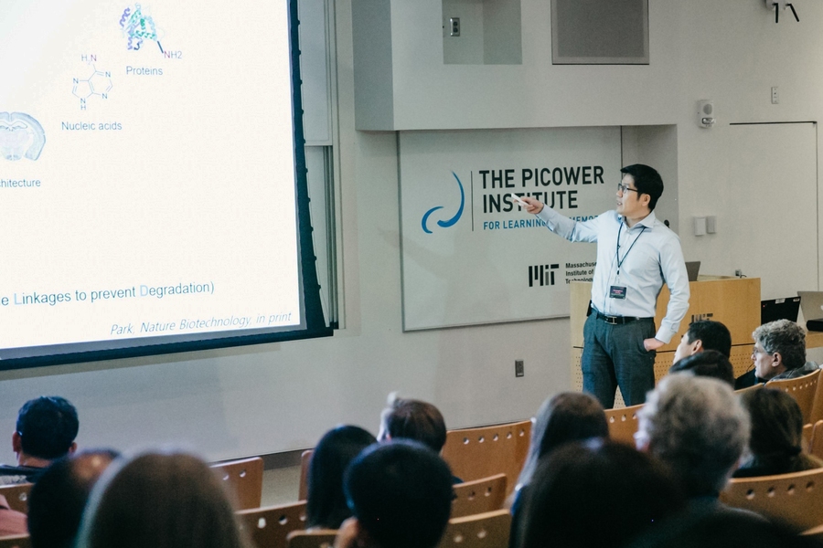
Hundreds of people who attended the Picower Institute for Learning and Memory fall symposium “Frontiers in Neurotechnology” on Oct. 23 got an inside look at emerging techniques and methods — ingenious new ways to look inside the brain, to understand its healthy anatomy and function, and to better understand disease.
Presented by 10 leading researchers the improvements in microscopy, advances in culturing brain tissues, and novel ways to detect and control brain activity illustrated the rapid pace of innovation in neuroscience today, said Li-Huei Tsai, Picower Professor and director of the Picower Institute. In her opening remarks, she reflected on how many new technologies her lab has adopted in the last decade and invited the crowd that packed Building 46’s Singleton Auditorium — and the overflow seating outside — to do the same.
“We are very fortunate to be working in neuroscience at a time of such extraordinary ingenuity,” she told the attendees.
Improving insight
Some of the new techniques that Tsai and hundreds of colleagues worldwide are adopting include methods of preserving, optically clearing, labeling, and enlarging brain tissue that were invented by the symposium’s host, Kwanghun Chung, assistant professor in the Picower Institute, the Institute for Medical Engineering and Science and the Department of Chemical Engineering.
At the symposium, Chung revealed that he is leading a new five-year project funded by the National Institutes of Health to map the entire human brain at unprecedented scales of detail, ranging from the circuits spanning distant regions down to individual synapses where neurons connect. The collaboration will take advantage of many of the rich suite of technologies his lab has developed. In recent work, Chung said, he’s also been applying the techniques to trace the circuits connecting brain regions that are key to deep-brain stimulation (a Parkinson’s disease treatment) and to illuminate differences between models of the autism-like condition Rett syndrome and healthy controls.
Advanced tissue processing, though, is just one category of ways that neuroscientists are getting a better look at the brain. Several speakers discussed major leaps in microscopy that have enabled the instruments able to image deeper, more clearly and faster, to keep pace with neural activity.
Elizabeth Hillman of Columbia University, for example, discussed her ongoing development of “SCAPE,” a version of light-sheet two-photon microscopy in which scopes image a broad plane of tissue rather than just a narrow spot. She showed how SCAPE is fast and sharp enough to allow real-time imaging of neural activity and blood flow in entire brains of behaving animals such as zebrafish and fruit flies, entire bodies of nematode worms, and large brain volumes in mice.
Na Ji of the University of California at Berkeley, meanwhile, described a different way of imaging activity within large brain volumes in live animals. She’s used the “Bessel beam” system to simultaneously image all the dendrites and synapses of a neuron (in 3-D) in the visual cortex to watch how it responds to specific sensory inputs. What would take 10 hours with a conventional scope takes 20 minutes using the technique, she said.
Ji and fellow speaker David Kleinfeld both pointed to another technology advancing microscopy: adaptive optics. This technology, already familiar to makers of advanced telescopes, dynamically adjusts the mirrors of a scope to mitigate distortions from light bending within complex tissues. By working to optimize parameters of two-photon microscopy, including the use of adaptive optics, Kleinfeld’s lab has been able to image 800 microns (0.8 mm) deep into the brain, which is enough to reach down to dendritic spines of neurons in layer 5 of the cortex, an important area of scientific interest because cells there receive input to those spines from the thalamus.
Beyond innovating upon and optimizing optics, another way speakers showed progress in seeing into the brain was in developing new kinds of “reporters,” or molecules that will fluoresce when they encounter a target chemical or protein. Kleinfeld, for example, has developed reporters called CNiFERs that show the concentration of neurotransmitters and neuromodulators. He’s used CNiFERs to show that mice can volitionally increase their levels of noradrenaline and dopamine when incentivized with rewarding feedback.
Boaz Levi of the Allen Institute discussed his work to develop reporters for an entirely different purpose: distinctly marking different cell classes and types in human and mouse cortex using engineered viruses. Even a rather specific region of the brain, after all, can have scores of different types of cells, each playing a diversity of roles. His goal is to help neuroscientists better sort through that complex landscape.
Yet another way to get a readout of brain activity is to monitor its large-scale oscillations, or brain waves. Though known for decades, brainwaves still have huge untapped potential to inform researchers and clinicians alike, said Emery N. Brown, an anesthesiologist-neuroscientist-statistician at the Picower Institute, Massachusetts General Hospital, and Harvard University.
By advancing rigorous statistical methods to analyze EEG readings of patients under general anesthesia, Brown has been able to create systems that allow for monitoring a patient’s brain state in real-time. The work has shed light on how anesthesia works, how people respond at different ages, and has led to innovations in medical practice that allow anesthesiologists to better control drug doses, often allowing for dramatic reductions in the amount of medicine administered, said Brown, the Edward Hood Taplin Professor at MIT. That, in turn, can help patients wake up faster and clearer-headed yet with better-managed pain.
The next step in Brown’s work, he said, is developing a real-time, closed loop system that can regulate dosing in response to EEG readings.
Making "mini-brains"
For ethical and logistical reasons, particularly when studying developmental, neurological and psychiatric disorders, scientists need to experiment with lab-grown neural tissue cultures rather than actual brains. Several speakers at the symposium shared their latest innovations in the burgeoning field of culturing 3-D brain organoids, or mini-brains, which can provide otherwise unattainable insights. Organoids are considered valuable because they can be grown from reprogrammed cells taken from human patients, or other organisms, and then developed into models that reflect brain development with those same genes. Moreover the genes can be precisely manipulated in the lab for experiments.
Sergiu Pasca of Stanford University, who published a review of how organoids can model various diseases the day after the symposium, offered several examples in his talk. For instance, in a new study his lab is examining the effects of oxygen deprivation on cells in the developing brain, a problem that sometimes affects fetuses.
Paola Arlotta of Harvard University also studies essential aspects of human brain development, such as the molecular rules that build neuronal diversity in the cerebral cortex, using organoid models. In her talk she emphasized the need to ensure that organoids provide reproducible experimental testbeds. After all, it’s one thing to grow a tissue but it’s another to make reliable experimental comparisons using them. She discussed work her lab has done to improve organoid culture to ensure greater reproducibility.
In a similar vein, Orly Reiner of the Weizmann Institute in Israel described how her desire to study a neurodevelopmental disorder using organoids motivated her to innovate to overcome challenges. Her lab is interested in understanding lissencephaly, a disease characterized by a lack of characteristic folds on the surface of the brain. But as organoids grow, if they don’t have vasculature, cells embedded deep inside can’t get nutrients they need and die. Moreover, without some of the kinds of innovations described above, microscopes can image those inner cells clearly. So Reiner’s lab decided to grow organoids on a chip, reshaping them into more of a thin pita bread-like shape that could be imaged and sustained. The models have indeed provided important insights into the disorder.
Arnold Kriegstein of the University of California at San Francisco has also used organoids to study lissencephaly and other disorders. His lab has a more general interest in a particular class of progenitor cells that give rise to neurons during development. He found that these progenitor cells behave abnormally in the lissencephaly model. Meanwhile, in a very different project, members of his lab are using organoids in comparative evolutionary biology studies that show key differences in brain development among humans, chimpanzees and macaque monkeys.
After a day filled with examples of neuroscientists describing new findings made possible by new technologies, Matthew Wilson, the Sherman Fairchild Professor in Neurobiology at MIT and associate director of the Picower Institute summed up the optimism of the field.
“This is the frontier,” Wilson said. “Thinking about these technologies that are giving us access to describe and understand the complexity of the brain at the level of molecules, cells and systems and then using that to turn around and understand how we can control brain function and understand cognition, it really is a golden age.”