
A new technique devised by MIT engineers may one day help physicians detect cancerous tumors during early stages of growth.
The technique allows nanoparticles to group together inside cancerous tumors, creating masses with enough of a magnetic signal to be detectable by a magnetic resonance imaging (MRI) machine.
The work appears as the cover feature in the May issue of Angewandte Chemie International Edition, one of the world's leading chemistry journals.
The research, which is just moving into animal testing, involves injecting nanoparticles (billionths of a meter in size) made of iron oxide into the body, where they flow through the bloodstream and enter tumors.
Solid tumors must form new blood vessels to grow. But because this growth is so rapid in cancerous tumors, there are gaps in the endothelial cells that line the inside of the blood vessels. The nanoparticles can slip through these gaps to enter the tumors.
Once inside the tumor, the nanoparticles can be triggered to group together by a mechanism designed by the MIT engineers. Specifically, certain enzymes, or proteases, inside the tumors cause the nanoparticles to "self-assemble" or stick together. The resulting nanoparticle clumps are too big to get back out of the gaps. Further, the clumps have a stronger magnetic signal than do individual nanoparticles, allowing detection by MRI.
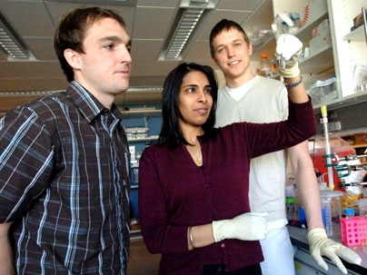
"We inject nanoparticles that will self-assemble when they are exposed to proteases inside of invasive tumors," said Sangeeta N. Bhatia, M.D., Ph.D., associate professor of the Harvard-MIT Division of Health Sciences & Technology (HST) and Electrical Engineering and Computer Science (EECS). "When they assemble they should get stuck inside the tumor and be more visible on an MRI. This might allow for noninvasive imaging of fast-growing cancer 'hot spots' in tumors." Bhatia also is affiliated with the MIT-Harvard Center of Cancer Nanotechnology Excellence.
The technique initially is being used to study breast tumors. Bhatia added that it eventually may be applied to many different types of cancers and to study the "triggers" that turn a benign mass in the body into a cancerous tumor. Nanoparticles also hold the promise of carrying medicines that could kill cancer cells, replacing radiation or chemotherapy treatments that cause negative side effects such as hair loss or nausea.
The researchers hold a provisional patent on their work.
Co-authors on the paper are Todd Harris and Geoffrey von Maltzahn, HST graduate students; Austin Derfus, a graduate student at the University of California at San Diego; and Erkki Ruoslahti, M.D., Ph.D., a professor at The Burnham Institute in LaJolla, Calif.
The work was supported by the National Cancer Institute, the National Aeronautics and Space Administration and the Whitaker Foundation.
A version of this article appeared in MIT Tech Talk on May 10, 2006 (download PDF).