Brain imaging methods allow scientists to peer into the human brain and observe the creation of new memories, with different forms of remembering emerging from different learning circuits, MIT neuroscientist Anthony Wagner reported at the AAAS meeting in Boston Feb. 14-19.
As illustrated in Christopher Nolan's movie "Memento," memories for our everyday experiences can be fleeting in the absence of brain mechanisms that transform our experiences into durable, long-term memory records. Studies of patients who suffer from anterograde amnesia, the condition that afflicted the central character in "Memento," are unable to lay down new long-term memories due to damage to structures in the inner (medial) portion of the temporal lobes (specifically, the hippocampus and the surrounding cortex).
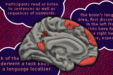
Wagner, the Paul E. Newton Assistant Professor of Neuroscience in the Department of Brain and Cognitive Sciences and the Center for Learning and Memory, said recent brain imaging data indicate that the medial temporal lobes do not act alone in building memories, but rather interact with the frontal lobes. He spoke on "Building and Retrieving Memories: Insights into the Cognitive Neuroscience of Memory" at a Feb. 16 AAAS symposium on "Images of Mind: Understanding How the Brain Enables Cognition."
The frontal lobes appear to play a role in allocating attention to different aspects of the experience, thus guiding the laying-down of memories that include these event details. Critical recent data suggest that the specific circuits engaged during memory formation depend on the nature of the experience (e.g., pictures of scenes vs. words), and how we attend to the experience (e.g., think about the meaning of the experience vs. think about its perceptual form).
These novel results from Wagner and colleagues provide important evidence regarding the organization of human memory and address the ongoing debate about how the brain acquires different kinds of new memories.
"Using this brain imaging technique that allowed us to characterize the human brain while it was in the process of building new memories, we gathered information on the moment when the brain gives birth to various forms of memory," Wagner said. "One set of results revealed a striking pattern: the neural processes that predicted whether someone would later remember or forget a visual experience were distinct from those that predicted whether he or she would later remember or forget a verbal experience. In particular, the left and right hemispheres of the frontal lobes played differentially greater roles in verbal and visual memory formation, respectively."
Keeping track of them
The main character in "Memento," Leonard, also illustrates that memories differ not only in their content but also in their detail. In his attempt to manage life in the face of his memory problem, he adopts a strategy that ultimately proves hazardous.
To remember key events, Leonard scribbles important information on snapshots that he keeps in his pocket or tattoos this information onto his body. These mementos--or memory records--provide him with some continuity across time and allow him to have a surrogate memory that records relevant facts about his past. By reading the notes on the photos or on his body, he can "remember" these facts.
However, one key component of memory that Leonard fails to record is the source of each fact--that is, from whom he learned the information and whether that source can be trusted. This renders him vulnerable to acting on information that he would otherwise discount.
This theme of "Memento" about the dangers of memory failure is summarized in a tattoo on Leonard's right bicep: "Consider the source--memory is treachery."
Recent data from Wagner and his colleagues suggest that these two aspects of memory for an experience--knowledge that one has encountered a stimulus before (item memory) and knowledge of the source or context of that experience--may depend on different learning mechanisms supported by distinct brain structures in the medial-temporal lobes.
Watching a memory form
Wagner and colleagues used a brain imaging technique called functional magnetic resonance imaging (fMRI) to collect images of the brain activity of healthy adults performing different memory tasks.
In all experiments, the participants were shown a long list of stimuli (e.g., words or pictures) and were instructed to attend to the stimuli during learning. While the participant was attending to the stimuli, fMRI recorded measures of neural activity from the individual's brain.
Following this learning task, the participants were removed from the fMRI scanner and, following a delay of between 20 minutes and 24 hours, their memory for the studied stimuli was then assessed. During this memory test, they were shown a long list of stimuli, some of which were new and some of which were presented to the participant while they were in the fMRI scanner. They were then asked to indicate whether they remember having seen the each stimulus during learning.
"This allowed us to assess various aspects of their memory for each event," Wagner said. "Most critically, based on their test responses we could sort the events encountered in the scanner into those that were later remembered and those that were later forgotten. Having this information, we could then ask what the brain was doing differently when it was building memories that ultimately could be remembered relative to when it was building memories that ultimately were forgotten."
The approach adopted by the researchers provides a unique window on the active brain and how differences in the brain's response during an experience relates to whether the experience will be remembered.
This work is funded by the National Institutes of Health, the Ellison Medical Foundation and the McKnight Fund for Neuroscience.
The BBC World Service did a radio piece on the work last weekend. Text and audio are available by clicking on the link at http://web.mit.edu/wagner/www/shameless.html.
According to reporter Caroline Ryan, "in addition to telling scientists more about how memory works, [Wagner's] technique could also one day be used to identify early signs of conditions such as Alzheimer's disease, where the brain's memory circuits stop working effectively."
A version of this article appeared in MIT Tech Talk on February 27, 2002.