CAMBRIDGE, Mass.--Contrary to popular belief, cells in the brain's primary visual cortex are "smart" enough to help determine where the eyes will look next, MIT researchers report in the June 13 issue of Science.
While the parietal and frontal cortex are thought of as key decision-makers that direct the gaze, "the primary visual cortex is much more than just a passive filter," said Mriganka Sur, head of MIT's Department of Brain and Cognitive Sciences and co-author of the study. "It is an active interpreter and creator of vision."
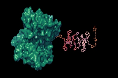
Discovering how internal representations of the world are created in our brains is regarded as central to understanding the neural basis of sensory and motor processing. "In this paper, we describe how the brain puts together an internal representation of the visual world," said Sur, a researcher in the Picower Center for Learning and Memory at MIT.
The primary visual cortex is where signals from the eyes are first processed. This brain area was long thought to be a filter that passed along information from incoming stimuli to other areas of the brain, but Sur and colleagues show that monkeys' primary visual cortex neurons are involved in high-level visual processing and decisions about where to look.
A Mind Picture
When we see, our brains create an internal representation of the visual world. This has to be updated constantly, incorporating new information. "If we understand how this image gets updated as a monkey acquires information about where it should look next, we can infer a great deal about how the brain works," Sur said.
In the primary visual cortex, cells respond to information streaming in from the millions of "wires" that relay sensory input from the eyes. The primary visual cortex takes a pixilated version of the world and creates one that highlights the edges of objects and compares intensities of light.
Still, if we could see what the primary visual cortex "sees" before it passes the image on to a higher functioning level of the brain, it would look like a very fragmented image.
Higher cortical areas also feed information back to the primary visual cortex. From this network of back-and-forth connections, the brain computes a crystal-clear Technicolor view of the world. The eyes scan the image as it is created, fixating on one part of the image at a time.
Until now, scientists considered eye movements and image analysis two largely separate parts of vision involving separate brain areas.
Seeing Spots
Co-author Jitendra Sharma, research scientist at the Picower Center, designed a simple experiment in which monkeys were shown a spot on a screen. The spot could appear at the center, right or left. With painstaking training, the monkeys learned to fixate on a spot for 2-3 seconds before receiving a reward.
A spot could appear randomly at any of the three locations or repeatedly at the same location. If the spot appeared in the same place more than once, the monkeys quickly caught on and anticipated the appearance of the spot. The study showed for the first time that neurons' responses to the same visual stimulus changed depending on an internal expectation of where stimuli would appear.
It turns out that the visual network of the brain works with almost clocklike predictability--you can apply the laws of probability to how neurons respond to an expected stimulus. "Past history of what you have just seen updates where you look next and how well you see," Sur said. The sharpness of the filter is determined by what has just been seen. "It's a dynamic filter at the very least -- a creative constructor of the image," he said.
Far from being a passive process, Sur is convinced that vision is influenced by hard-to-quantify entities such as attention, expectation and motivation. To pick apart these elements of cognition, Sur is examining the interactions between vision and these internal states of mind.
In addition to Sur and Sharma, authors include Valentin Dragoi, postdoctoral fellow at the Picower Center; Joshua B. Tenenbaum, assistant professor in brain and cognitive sciences; and Earl K. Miller, professor of brain and cognitive sciences and associate director of the Picower Center.
This work was supported by the National Institutes of Health.