For the first time, MIT researchers have identified a gene involved in the retrieval of long-term memories. The study will appear in the May 30 online edition of Science.
Nobel laureate Susumu Tonegawa, director of the Picower Center for Learning and Memory at MIT and the Picower Professor of Biology and Neuroscience, said his latest discovery may lead to drugs that would counter the memory loss that plagues Alzheimer's victims and relieve middle-agers' "senior moments."
Studies have shown that older people have not lost their memories--they just have trouble retrieving them, Tonegawa said. Similarly, the genetically altered mice in the MIT study have no trouble forming a long-term memory of a specific place, but they cannot retrieve the information with a limited number of cues.
This process, called pattern completion, is believed to be a crucial step in the retrieval of memories. "In real life, we always recall memories with limited information," he said. "A memory is dormant until it is retrieved from the subconscious to the conscious with cues."
Tonegawa's laboratory explores the molecular, cellular and network mechanisms underlying memory. He has pioneered the development of a genetic technology that permits researchers to create a new strain of mice in which a specific gene is knocked out in a specific area of the brain. In previous studies, Tonegawa and colleagues at MIT used the genetically engineered mice to show that a gene for the NMDA receptor in a region of the hippocampus called CA1 plays a crucial role in the formation of a long-term memory.
For these studies, Tonegawa generated and analyzed mice with a genetically altered hippocampus, a brain structure that has been shown to play a crucial role in common types of memories.
In addition to Tonegawa, the paper's authors are Matthew A. Wilson, associate professor of brain and cognitive sciences; Kazutoshi Nakazawa, research scientist in the Picower Center for Learning and Memory; graduate students Michael C. Quirk and Linus D. Sun; research associate Candice A. Carr; MIT affiliate Akira Kato; and others from the Baylor College of Medicine and the Hokkaido University School of Medicine.
FORMING MEMORIES
The formation of a long-term memory starts with input through one of the five senses. We store an endless array of information as memories. Much of it is tucked away in the brain's cellular networks at the subconscious level. To recall a memory, we need cues to activate the memory network and bring the stored information to the conscious level.
"Very limited cues are sufficient to trigger a chain reaction that permits us to become aware of the rich and detailed content of a memory," Tonegawa said. "This phenomenon is called pattern completion because it reflects cellular processes accompanying memory retrieval in which reactivation of a pattern of cellular connections harboring memory is completed by very limited input."
The hippocampus is divided into three sections. Tonegawa looked at cells in the CA3 region, in which individual cells work in a so-called auto-association network by sending signals back to each other as well as forwarding them to other cells. Mathematical models suggest that through the modification of cellular connections, memories stored in such a network can be efficiently accessed for the purpose of pattern completion. Activity passed between cells through these connections can replace information that might be missing in the input. In this way, the network is able to "fill in" or retrieve complete memories given only a subset of the original cues that were present when it was formed.
"When people get old, they complain they're losing their memory. In fact, in many cases they are losing their ability to recall memories," Tonegawa said. "It's the tip-of-the-tongue phenomenon--if they had more triggers, they often can recall the facts." The same is true in the early phase of Alzheimer's disease. "It's not that they lost the memory. It's that they have a retrieval problem," he said.
MICE AND MEMORY CUES
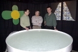
In the experiment, mice were given a learning and memory challenge: locate a submerged platform hidden from view in a pool of water.
Although mice can swim, they don't like water, so they immediately searched for a way out. After swimming around randomly, they would find the platform and climb out. Visual cues placed around the tank helped the mice orient themselves.
Each mouse that repeated the task several times a day for a week got better at finding the platform, until they got to a point where they swam straight to it. After the animal had learned the task, several of the visual cues outside the tank were removed.
Even with only one visual cue remaining, normal mice remembered the location of the platform quickly and easily. But with only one cue, mice with altered NMDA receptors in the CA3 area of the hippocampus could not find the platform. They could only find it with all four cues intact.
This study is striking because it provides evidence at the behavioral level as well as the molecular level, Tonegawa said. To provide insight into the link between these levels, Wilson has devised a method to monitor the actions of dozens of individual neurons in real time.
With electrodes that reach into the depths of the hippocampus, the researchers can actually see memories being formed in the brains of their mice subjects. Each memory creates a unique pattern of activity in the hippocampus. Individual cells called place cells fire in response to memory activation of different places.
Using these recording techniques, the researchers were able to identify the characteristics of neural activity corresponding to the partially cued reactivation of previously formed memories, or pattern completion.
When animals explored an environment in which all four visual cues were present, the patterns of place cell activity in the genetically altered mice appeared to be normal.
But when cues were removed, the cells responded much more weakly than cells in normal animals. The weakened response was attributed to the inability of the CA3 area to successfully retrieve the complete memory of that environment given only partial cues.
"This result provides a direct measurement of how failed memory retrieval might appear at the level of individual neurons, and therefore provide the basis for improving memory under these conditions," Wilson said.
The NMDA receptor is a receptor for the neurotransmitter glutamate, which is the most important excitatory transmitter in the brain. It is not only a receptor but also a channel that seems to be involved in many processes such as synaptic plasticity and target recognition.
The density of NMDA receptors is reduced in older people. The CA3 region of the hippocampus is known to be most vulnerable to plaque formation in the brains of Alzheimer's patients.
"By understanding how memory retrieval works at the molecular level, this could be the basis for drug development in the future," Tonegawa said. "It's important not only for Alzheimer's patients, but for prolonging the ability of aging people to recall memories and learned facts."
This work is supported by the National Institutes of Health, RIKEN, the Howard Hughes Medical Institute and the Human Frontier Science Program.
A version of this article appeared in MIT Tech Talk on June 5, 2002.


