The Athinoula A. Martinos Center for Biomedical Imaging dedicated two state-of-the-art imaging systems Monday. The systems are the first of their kind in the Northeast.
A new 7-Tesla magnetic resonance imaging system will permit researchers to image the brain with sufficient resolution to identify tiny anatomic details and to localize regions important for specific brain functions such as memory, language and reward.
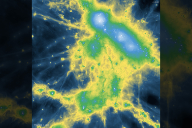
The second system, a 306-channel magnetoencephalograph, will image the brain's electrical activities with very high-temporal resolution in real time. Using scientific approaches pioneered by researchers at the Athinoula A. Martinos Center, the combined MEG and MRI data from these new facilities will produce "movies" of the brain at work, thus providing a fresh view of how different parts of the brain communicate with each other.
"We are deeply grateful to the Office of National Drug Control Policy , the Mental Illness and Neuroscience Discovery Institute , the Congress--particularly senators Peter Domenici from New Mexico and Edward Kennedy from Massachusetts--and the American people for the extraordinary support that they have provided the Athinoula A. Martinos Center for Biomedical Imaging," said Bruce R. Rosen, director of the center and a senior lecturer in nuclear engineering at MIT. "We share their dedication to better understanding the workings of the human brain, and to translating that knowledge into improved treatments and, ultimately, the prevention of substance abuse and mental illness."
The Martinos Center in Charlestown, Mass. is a collaboration among MIT, Massachusetts General Hospital and Harvard Medical School through the Harvard-MIT Division of Health Science and Technology and the Massachusetts General Hospital's Department of Radiology. The center's mission is to create, develop and apply innovative and advanced imaging technologies that permit a more comprehensive understanding of, and better care for, the human mind and body.
Research at the center is focused on nuclear magnetic resonance technology--from hardware and software development to a range of imaging applications, to advanced data post processing, modeling and statistics.
Using high-speed and high-field magnetic resonance (MR) imaging, MR spectroscopy, optical imaging, magnetoencephalography (MEG), and electroencephalography (EEG), Martinos investigators explore the properties of biological systems to help understand and develop new treatments for human pathologies such as cancer and neurological and cardiovascular disorders.
The development and application of novel imaging technologies go hand-in-hand at the center. New technologies provide insights and opportunities to study human disease and biology in ways previously not possible. At the same time, the scientific questions and goals of those studying a particular disorder or phenomenon drive the development of new technologies. Among the MIT researchers who have expressed interest in using the new MEG system for their own work are Anthony Wagner, the Paul E. Newton Career Development Assistant Professor in Neuroscience; Nancy Kanwisher, professor of brain and cognitive sciences; and Suzanne Corkin, professor of behavioral neuroscience.
HELP WITH WAR ON DRUGS
The new 7-T MRI system, made possible through a grant from the Office of National Drug Control Policy (ONDCP), will help researchers understand the biological basis of addiction and will contribute to developing better treatments and deterrents for addiction.
Using this system, researchers can look at biochemistry and metabolism in the brain, and measure neurochemicals with greater precision than ever before. It will improve understanding of the brain's circuitry and how that circuitry differs in patients who suffer from substance abuse or other neurological or psychiatric conditions, including Parkinson's disease, Alzheimer's disease, schizophrenia, depression and chronic pain.
This work will allow scientists to develop a more comprehensive perspective on how drug abuse captures and changes brain function. Previously, researchers at the Martinos Center had demonstrated the commonality of the brain's circuitry involved in drug euphoria and craving, pain, gambling and social rewards.
"A key element of the federal government's war on drugs is to work with the best equipment and researchers in the country to advance our understanding of the biological basis of addiction. We are delighted to be here today at the Athinoula A. Martinos Center for Biomedical Imaging to dedicate this new state-of-the-art MRI system," said Al Brandenstein, director of the ONDCP's Counterdrug Technology Assessment Center, at the dedication ceremonies.
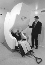
The newly installed MRI system contains a 7-Tesla, 90-cm whole-body magnet built by Magnex Scientific and a 460-ton steel shield. The console, gradient drivers and patient table are provided by Siemens Medical Systems.
The integration of its components, as well as the design and construction of its radio-frequency coils, was performed by MGH personnel. Because the ultrahigh-field magnet is fully integrated into a conventional MRI console, the 7-T system will enable investigators to apply their scientific findings to improve patient care on more widely available clinical MRI systems.
SYSTEM HAS MIT ROOTS
The second newly installed facility, a MEG system, was acquired through a partnership with the Mental Illness and Neuroscience Discovery (MIND) Institute, based at the University of New Mexico. MIND's mission is to explore the mind and brain to enhance the lives of individuals with mental illness. MIND advances and applies neuroimaging technologies to bridge the emerging frontiers of basic neuroscience and their clinical applications.
"In order to improve the lives of patients with mental illness, and their families, it is critical to support initiatives that can shed light on the neurobiological basis of mental illness. We believe this MEG system will provide researchers with a dynamic new tool with which to explore and understand better the workings of the brain," said Nancy Andreasen, director of the MIND Institute.
The new MEG system features a 4-D Neuroimaging/Neuromag Vectorview 306-channel whole-head neuromagnetometer. More than 128 channels of EEG can be simultaneously recorded.
The MEG/EEG laboratory is considered very quiet for the facility's urban location, and the magnetically shielded room is one of the most effective in the world. The room is constructed with three thick layers of a special magnetically shielded iron. The laboratory is equipped with complete auditory, visual and somatosensory stimulation systems. The research suite also includes a whole-head, combined 64-channel EEG; advanced imaging systems that use near-infrared light for diffuse optical tomography and appropriate behavioral stimulation devices.
"The new MEG system allows MIT researchers to work at the cutting edge of this technology," said David Cohen, a pioneer in the field of magnetoencephalography who helped design the MEG system and the shielded room that houses it. "Not only is the MEG equipment state-of-the-art, but the fact that it is located in the Martinos Center allows the MEG to be combined with MRI, fMRI and other forms of brain imaging. This is the very new multi-modal approach."
The first MEG system using a shielded room was operated at MIT's Francis Bitter Magnet Laboratory for many years under Cohen's direction. Cohen, now a group leader at the Magnet Lab who is considered the inventor of the MEG system, first measured the magnetoencephalogram in 1968. He also works at the Martinos Center and is planning an experiment on the new MEG system.
At the dedication, a two-foot-wide scale model was displayed of Cohen's original MIT room where the MEG was born. "It shows that the MEG indeed started at MIT," Cohen said.
The Martinos Center has more than 40 faculty members and 80 postdoctoral and graduate students with expertise in various disciplines, including the engineering and physical sciences, computational sciences and informatics, behavioral and cognitive neurosciences, basic and applied biological sciences, imaging and radiological sciences, and clinical sciences.
The center also is a resource to biomedical imaging researchers from institutions in the Boston area and from around the world. A vital component of the center's research is its partnerships with government, industry and private foundations, including the National Center for Research Resources, the Office of National Drug Control Policy, Siemens and the MIND Institute.
A version of this article appeared in MIT Tech Talk on May 1, 2002.