CAMBRIDGE, Mass. -- Researchers at the Massachusetts Institute of Technology and the University of Wales report in the Sept. 28 issue of Science that they have identified a part of the brain that responds primarily to images of the human body or parts of the body.
This is the latest in a series of findings on highly specific brain functions by Nancy Kanwisher, professor of brain and cognitive sciences at MIT. With the help of state-of-the-art brain imaging, Kanwisher and colleagues had previously uncovered the parahippocampal place area, which responds to photographs of indoor and outdoor scenes, and the fusiform face area, which responds exclusively to faces.
The newest region is on the lateral surface of the brain, just inside the skull from a point behind the ears. The paper's authors, who include Paul E. Downing of the Centre for Cognitive Neuroscience at the University of Wales, MIT postdoctoral fellow Yuhong Jiang and research assistant Miles M. Shuman, named it the extrastriate body area (EBA).
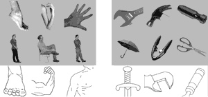
The EBA responds strongly to pictures eyes, feet, hands, ears and elbows, as well as to photos, silhouettes, artists' renderings and stick figures of entire humans, and responded much less strongly to similarly shaped inanimate objects and stick figures that had been rearranged so they no longer had a human shape. It responded moderately to pictures of mammals and no more to a human face than to an inanimate object.
FACES, PLACES AND MORE
"We have been looking for brain regions that exhibit any kind of selective response," Kanwisher said. "We had found selective neural responses to faces and places and we wondered, 'What else?' We tested a number of different categories, but we never expected to find this," although there are a few reported cases of people who, after suffering brain damage, lose the ability to name parts of the body.
The finding opens up a host of questions. Why do we have cortical regions that are preferentially involved in processing faces, places, and bodies, but apparently not spiders, cars or food? And what exactly does the EBA do with images of bodies? The researchers speculate that it may help identify individuals when the face is hard to see, or determine the body configuration of another person or help us place our own body parts in space, but further research is needed.
"This is an exciting time to be working in the field of cognitive neuroscience," Kanwisher said. "Thanks in large part to fMRI (functional magnetic resonance imaging, a harmless technique to study the brains of normal subjects), scientists are now making progress at a historically unprecedented rate in understanding the functional organization of the mind and brain -- including the discovery of what Noam Chomsky once called the 'organs' of the mind."
A SERIES OF SPECIALTIES
Neuroscientists are now seeking clues to whether the human mind consists of a single, multi-purpose system or a constellation of distinct, independent systems, each specialized for a particular mental task.
Evidence comes in part from the striking abnormalities exhibited by victims of brain damage. Prosopagnosics, for example, cannot recognize faces, including their own.
The use of fMRI has already turned up a number of brain regions with stunningly specific functions: face and place identification.
"We wanted to know just how many of these category-specific regions existed in the human brain. To find out, we used fMRI to scan the visual areas of the brains of volunteers while they viewed pictures of objects from over a dozen different categories such as food, animals, tools, faces, musical instruments and human body parts," Kanwisher said.
"We looked for areas of the brain where neural activity was consistently higher. Our search turned up very little evidence for new brain areas that responded selectively to any of the new categories tested, except for one. In every subject tested, a region on the lateral surface of the brain, just inside the skull, responded more strongly when subjects viewed photos of human body parts than when they viewed any of the other categories of objects," she said.
To make sure that this region was not responding to something such as the presence of smooth, curved surfaces, the researchers ran additional experiments. The brain's "body area" consistently produced a stronger response to line drawings of bodies and parts of the body than to line drawings of objects and object parts.
The researchers also ruled out the possibilities that the EBA was responding to anything living, anything known to be capable of motion or any object with parts that can move relative to each other. The region responded more to human bodies than to trees, mammals, cars or objects with movable parts like scissors, staplers and corkscrews.
WHAT DOES IT MEAN?
The EBA is in a region of the brain called the posterior superior temporal sulcus, where other areas have been shown to be activated during social perception--the perception of socially relevant information such as the direction that another person's eyes are gazing, the sound of human voices or the inferred intentions of animate entities.
The EBA may be part of a broader network of nearby areas involved in social perception and social cognition.
"Although the discovery of this new area is exciting, we are only now at square one, with the most important questions yet to be answered.
"Beyond determining the function of this area, it will be important to find out how it originates in the cortex in the first place. Does the genome specify that this part of the brain is destined to process information about human bodies? Or does the specialization of this region result from a self-organizing process whereby visual functions become segregated in the cortex as a function of each individual's perceptual experience?" Kanwisher said.
This work is supported by a grant from Human Frontiers.