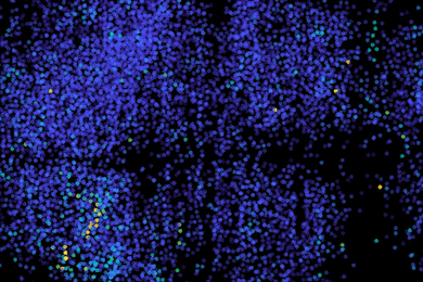
High-speed microscopy reveals electrical activity across the brain
The new technique could help scientists learn how the entire brain works to generate decisions and emotions.
The new technique could help scientists learn how the entire brain works to generate decisions and emotions.
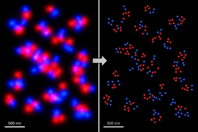
New U-STORM imaging technology lets scientists view molecular structures in subatomic detail — about 1,000 times clearer than traditional dyes — while making the microscope process much simpler.
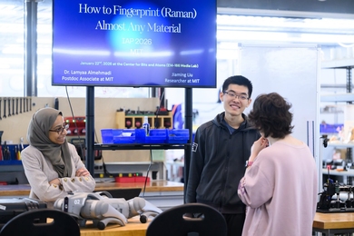
Participants learn how laser “fingerprinting” can help identify materials in fields ranging from law enforcement to art restoration.

The method enhances 3D bioprinting capabilities, accelerating process optimization for real-world applications in tissue engineering.
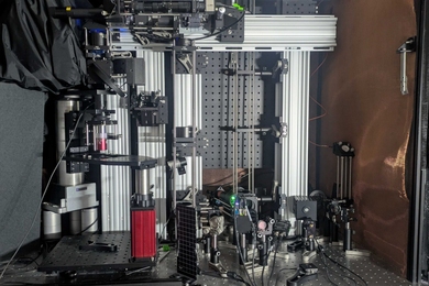
By combining several cutting-edge imaging technologies, a new microscope system could enable unprecedentedly deep and precise visualization of metabolic and neuronal activity, potentially even in humans.
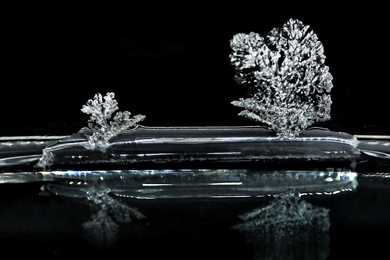
Device Research Lab study uncovers mechanisms behind a phenomenon that can impact civil engineering, desalination, coatings, membrane design, art conservation, and more.

Since an MIT team introduced expansion microscopy in 2015, the technique has powered the science behind kidney disease, plant seeds, the microbiome, Alzheimer’s, viruses, and more.
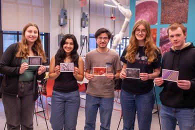
The undergraduate lab’s first microscope competition highlights stunning images and student ingenuity.
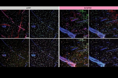
New methods light up lipid membranes and let researchers see sets of proteins inside cells with high resolution.
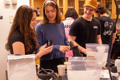
Undergraduate class blends science, hands-on experimentation, and a love for coffee to fuel curiosity.
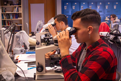
A summer class teaches PhD students and early-career archaeologists ceramic petrography, revealing the origins and production methods of past societies.
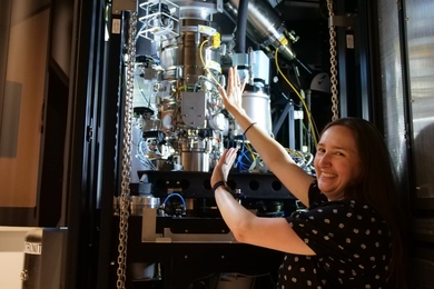
For Sarah Sterling, the new director of the Cryo-Electron Microscopy facility at MIT.nano, better planning and more communication leads to better science.

A trailblazer in electron microscopy, Vander Sande is remembered for his dedication to teaching, service, and global collaboration.
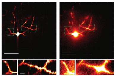
A newly described technology improves the clarity and speed of using two-photon microscopy to image synapses in the living brain.
Physics professor will use the award to develop a new kind of microscopy.