For the human brain, birth is a great divide. Like marble ready for sculpting, the prenatal brain abounds in extraneous neurons and connections waiting for experiences to carve the neural circuits that enable us to perceive, think and learn.
If this sculpting, known as plasticity, goes awry in early development, neurological disorders can result. Even certain late onset conditions like schizophrenia, Huntington's and Lou Gehrig's disease may have origins in poor wiring that occurs very early, at a time that is largely ignored in disease targeted research.
"Until this study, we did not realize how profoundly the process of wiring those circuits differs in early development from later stages," said MIT Professor Martha Constantine-Paton of biology and the McGovern Institute for Brain Research at MIT. Her paper appeared in the Dec. 22 online edition of the Journal of Comparative Neurology, in advance of its Feb. 10 publication.
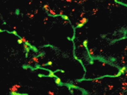
Her study focused on the visual system in rats, a convenient model for studying plasticity and how new circuits develop because retinal neurons are just one synapse away from the brain. Retinal cells must map to a specific location in a portion of the rat's brain called the superior colliculus.
Most researchers study synapse number and turnover in juvenile and adult animals after eye opening, analogous to birth in humans. At that stage, the brain strengthens selected axon synapses that correctly reach and enervate their target cells.
No one had actually studied these processes in neonate animals prior to eye opening, a period analogous to prenatal humans. But most scientists assumed it worked about the same as after the onset of vision -- a faulty assumption, the McGovern researchers found. Instead of strengthening the right connections, the brain simply eliminates the weak synapses connected to the wrong place prior to eye opening.
"It's similar to choosing players for a young Little League versus a professional team," explains first author Matthew Colonnese, a former postdoctoral researcher in Constantine-Paton's lab who is currently conducting research in the lab of Alan Jasanoff, an associate member of the McGovern Institute at MIT. "Most very young players are not very good, so coaches take all comers and then quickly weed the weaker players from first string. But pro coaches recruit proven players, so fewer need weeding out."
This Little League strategy probably happens because before eye opening, the retinal neurons fire spontaneously. But the brain cannot know which connections to strengthen because they have not yet responded to patterns of light. So it simply eliminates grossly misguided and relatively ineffective axons.
At both Little League and pro levels, the "coach" is the NMDA receptor, a protein that responds to the excitatory signal glutamate. Scientists have known that later in life, this receptor acts like the pro coach, mostly strengthening existing axon connections. Few anticipated the very robust role it plays in the Little League, before eye opening, when the receptor exclusively eliminates the weaker players.
Yet that role makes sense, suggests Colonnese. Otherwise, the brain would have too many axons and a chaotic neural circuitry. He predicts that researchers will probably discover many other ways in which the outcome of NMDA receptor function differs in fetuses and children versus teenagers and adults.
This research was supported by the National Institutes of Health.