CAMBRIDGE, Mass. -- A $20 million gift from a Greek couple will create a new center within the Harvard-MIT Division of Health Sciences and Technology (HST) dedicated to biomedical imaging.
MIT President Charles M. Vest and Joseph B. Martin, dean of Harvard Medical School, announced that the gift from Thanassis and Marina Martinos of Athens will establish the Athinoula A. Martinos Center for Functional and Structural Biomedical Imaging, named for the couple's late daughter.
Biomedical imaging, a relatively young field, enables physicians and scientists to "see" and better understand tissue and organ function. It provides physicians with the ability to visualize the structure of tissues and to capture their function on film.
HST brings together science and engineering at MIT and Harvard, the Harvard Medical School, and its affiliated hospitals and research centers to solve problems in human health. The largest biomedical engineering and physician scientist training program in the United States, HST is responsible for significant advances in biology, health-related technology and medicine.
"The new center is a profoundly important venture that, taken together with our collaborating clinical imaging centers, can dramatically improve the approach to scientific inquiry and clinical management. This center, to be located on the MIT campus, will help define the future of imaging well into the next century," Vest said.
Thanassis Martinos said, "It is a great privilege to support the Harvard-MIT Division of Health Sciences and Technology in this ambitious effort. Our goal is to make a meaningful contribution that will advance our understanding and treatment of disease." Martinos and his wife have the controlling interest in Eastern Mediterranean Maritime, a major shipping company, as well as significant real estate holdings in Greece.
"SEEING" INSIDE THE BODY
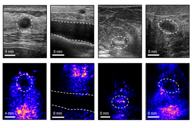
One type of biomedical imaging is magnetic resonance imaging (MRI), also known as nuclear magnetic resonance (NMR). Using electromagnetic fields and radio waves to read minute shifts in the magnetic alignment of protons in soft tissue such as the brain, it involves the collaboration of engineers, computer scientists, neuroscientists and physicians.
The important advance called functional MRI (fMRI) shows how living tissues are functioning in real time. For example, fMRI can make a 100-millisecond scan every few seconds to detect variations in regional blood flow within the brain to signal sight, hearing, thinking or feeling. Combining many fMRI scans makes a real-time "movie" of functioning organs that works like a flip-book. This breakthrough has been especially useful in cognitive neuroscience and psychology.
Current imaging research at HST that will be advanced by the Martinoses' gift includes:
NMR brain imaging, which helps physicians determine how best to save portions of the brain at high risk of damage from stroke or disease. A wide range of measurements of brain function has provided new ways to monitor experimental therapies and has allowed an unprecedented degree of rehabilitation to stroke patients.
This work at the Massachusetts General Hospital (MGH) NMR Center in Boston is headed by HST graduate and faculty member Dr. Bruce R. Rosen.
Video-guided imaging, a technique used by neuro surgeons, provides a detailed picture of the brain superimposed on the patient's skull, offering surgeons a previously unimaginable level of precision and detail.
With the system, a surgeon can tell the exact location of structures such as critical blood vessels and tumors. Because the video is live, the surgeon is able to watch his or her own hand on the monitor. As a result, the surgeon knows precisely where to make cuts.
At the heart of the system is software that allows precise alignment of images. "Our algorithm gives us a totally automatic way of taking a view of a patient, and taking a model of the 3D internal anatomy of that patient, and exactly lining them up," said Dr. Eric Grimson, an HST-MIT faculty member who leads this team in the Surgical Planning Laboratory of Boston's Brigham & Women's Hospital.
A LONG-STANDING RELATIONSHIP
The Martinoses' connection to HST goes back more than 20 years. In 1976, on a Friday afternoon, Martinos tracked down Dr. Daniel C. Shannon, who was at that time director of Pediatric Intensive Care at MGH and a member of HST's founding faculty.
Martinos asked Shannon, who was on vacation in Nantucket, to come to Greece to treat a desperately ill godchild. Shannon arrived in Athens on Saturday and, unable to identify the cause of heart and lung failure, as well as coma, returned to Boston with the child the following day, risking 10-1 odds against the child's surviving a flight to Boston. She was admitted to the MGH Pediatric Intensive Care Unit for diagnosis and treatment, and within two weeks, the child was back in Greece. Today she is 26 years old and a graduate student. Thus began a long and deep friendship between Shannon and the Martinoses.
When their oldest daughter, Athinoula, died in 1997 at age 24, the Martinoses spoke with Shannon, who also had lost a daughter, about how he had dealt with his loss. One way Shannon coped was to establish a research scholarship fund for young women at his daughter's college. Shannon suggested that the Martinoses set up the Athinoula A. Martinos Research Scholarship fund to support the research, study and training of HST students. The first 10 scholars in this on-going program were announced at the 1997 HST Research Forum at MIT.
ADVANCING TREATMENT AND KNOWLEDGE
Shannon and HST co-directors Dr. Martha L. Gray and Dr. Joseph V. Bonventre focused the Martinoses' recent attention on biomedical imaging, knowing the couple is interested in supporting work that could advance medical knowledge and develop innovative treatments for brain disease.
"The Martinos Imaging Center will be an important physical representation of HST's commitment to the solution of biomedical problems and improvement of human health by advancing imaging technologies that by their very nature integrate scientific and medical disciplines. We applaud the members of the Martinos family for their vision of the future of research at the interface of science, technology and medicine," Bonventre said.
"Imaging is still a young science," Gray said. "With the scientific and technological strengths of MIT, blended with the clinical strengths of Harvard Medical School and its affiliated teaching hospitals in Boston, we are confident that tremendous strides can be made in advancing this valuable technology."