


Do a Google search for dark-field images, and you’ll discover a beautifully detailed world of microscopic organisms set in bright contrast to their midnight-black backdrops. Dark-field microscopy can reveal intricate details of translucent cells and aquatic organisms, as well as faceted diamonds and other precious stones that would otherwise appear very faint or even invisible under a typical bright-field microscope.
Scientists generate dark-field images by fitting standard microscopes with often costly components to illumate the sample stage with a hollow, highly angled cone of light. When a translucent sample is placed under a dark-field microscope, the cone of light scatters off the sample’s features to create an image of the sample on the microscope’s camera, in bright contrast to the dark background.
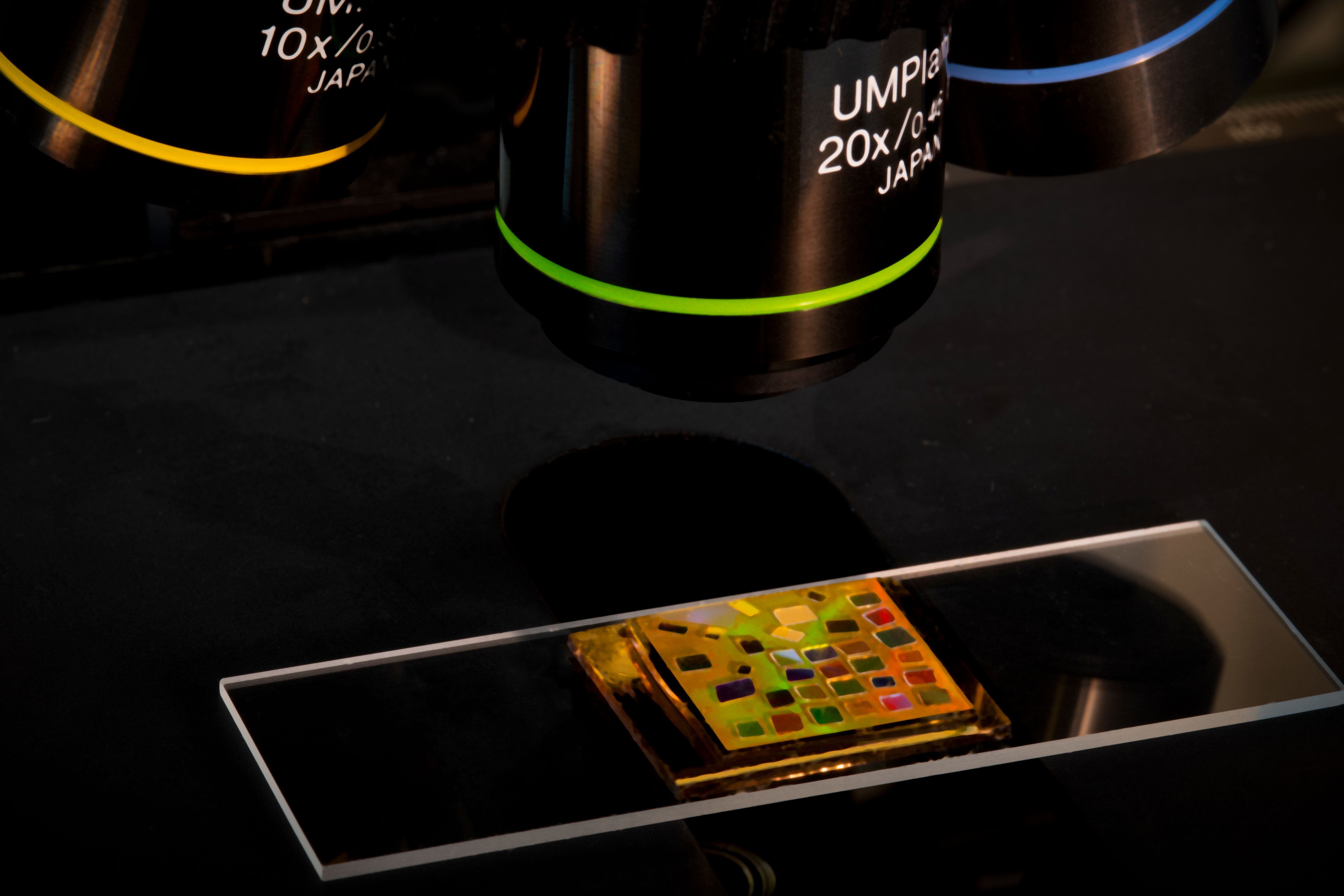
Now, engineers at MIT have developed a small, mirrored chip that helps to produce dark-field images, without dedicated expensive components. The chip is slightly larger than a postage stamp and as thin as a credit card. When placed on a microscope’s stage, the chip emits a hollow cone of light that can be used to generate detailed dark-field images of algae, bacteria, and similarly translucent tiny objects.
Credit: Cecile Chazot
The new optical chip can be added to standard microscopes as an affordable, downsized alternative to conventional dark-field components. The chip may also be fitted into hand-held microscopes to produce images of microorganisms in the field.
“Imagine you’re a marine biologist,” says Cecile Chazot, a graduate student in MIT’s Department of Materials Science and Engineering. “You normally have to bring a big bucket of water into the lab to analyze. If the sample is bad, you have to go back out to collect more samples. If you have a hand-held, dark-field microscope, you can check a drop in your bucket while you’re out at sea, to see if you can go home or if you need a new bucket.”
Chazot is the lead author of a paper detailing the team’s new design, published today in the journal Nature Photonics. Her co-authors are Sara Nagelberg, Igor Coropceanu, Kurt Broderick, Yunjo Kim, Moungi Bawendi, Peter So, and Mathias Kolle of MIT, along with Christopher Rowlands at Imperial College London and Maik Scherer of Papierfabrik Louisenthal GmbH in Germany.
Forever fluorescent
In an ongoing effort, members of Kolle’s lab are designing materials and devices that exhibit long-lasting “structural colors” that do not rely on dyes or pigmentation. Instead, they employ nano- and microscale structures that reflect and scatter light much like tiny prisms or soap bubbles. They can therefore appear to change colors depending on how their structures are arranged or manipulated.
Structural color can be seen in the iridescent wings of beetles and butterflies, the feathers of birds, as well as fish scales and some flower petals. Inspired by examples of structural color in nature, Kolle has been investigating various ways to manipulate light from a microscopic, structural perspective.
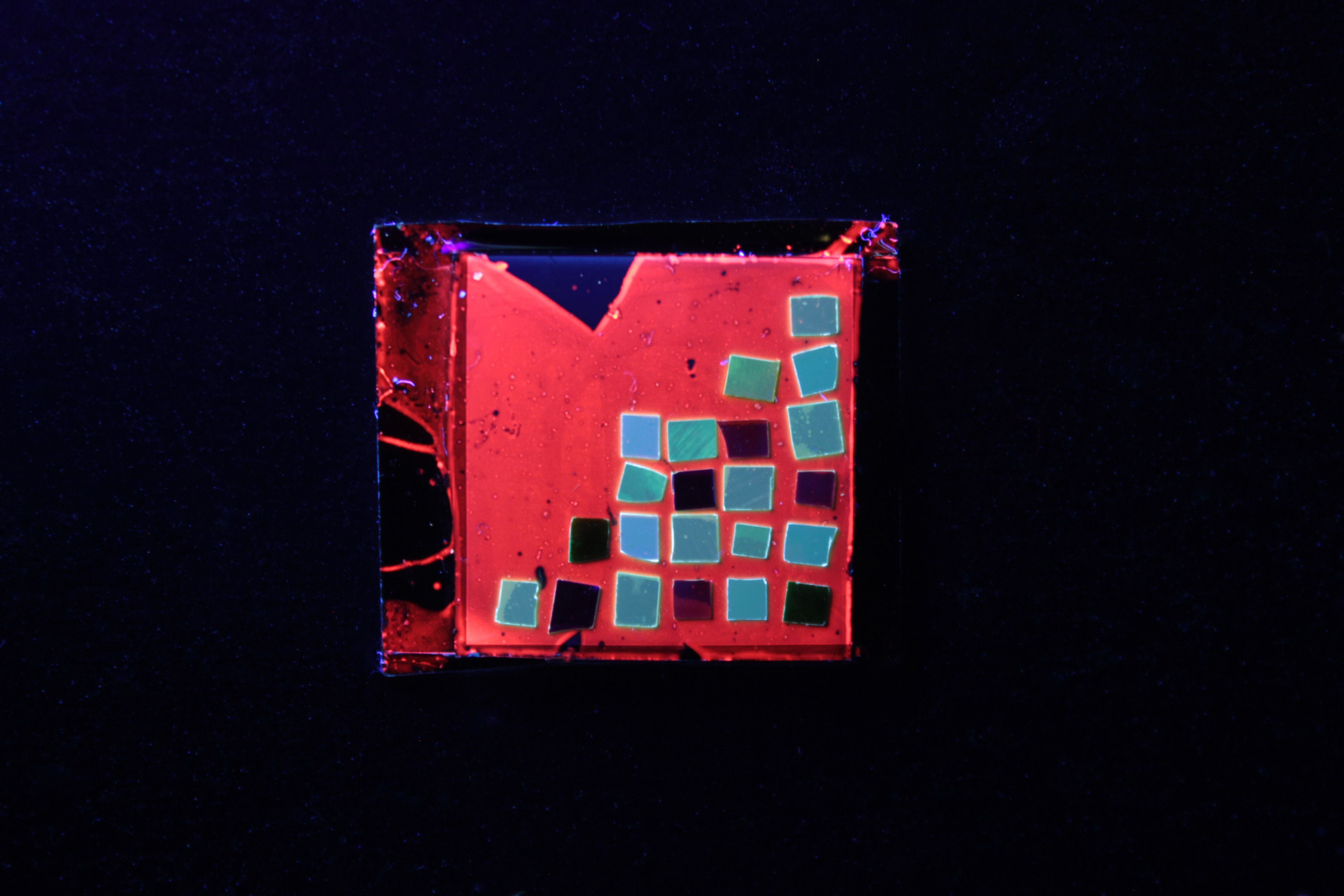
As part of this effort, he and Chazot designed a small, three-layered chip that they originally intended to use as a miniature laser. The middle layer functions as the chip’s light source, made from a polymer infused with quantum dots — tiny nanoparticles that emit light when excited with fluorescent light. Chazot likens this layer to a glowstick bracelet, where the reaction of two chemicals creates the light; except here no chemical reaction is needed — just a bit of blue light will make the quantum dots shine in bright orange and red colors.
“In glowsticks, eventually these chemicals stop emitting light,” Chazot says. “But quantum dots are stable. If you were to make a bracelet with quantum dots, they would be fluorescent for a very long time.”
Over this light-generating layer, the researchers placed a Bragg mirror — a structure made from alternating nanoscale layers of transparent materials, with distinctly different refractive indices, meaning the degrees to which the layers reflect incoming light.
The Bragg mirror, Kolle says, acts as a sort of “gatekeeper” for the photons that are emitted by the quantum dots. The arrangement and thicknesses of the mirror’s layers is such that it lets photons escape up and out of the chip, but only if the light arrives at the mirror at high angles. Light arriving at lower angles is bounced back down into the chip.
The researchers added a third feature below the light-generating layer to recycle the photons initially rejected by the Bragg mirror. This third layer is molded out of solid, transparent epoxy coated with a reflective gold film and resembles a miniature egg crate, pocked with small wells, each measuring about 4 microns in diameter.
Chazot lined this surface with a thin layer of highly reflective gold — an optical arrangement that acts to catch any light that reflects back down from the Bragg mirror, and ping-pong that light back up, likely at a new angle that the mirror would let through. The design for this third layer was inspired by the microscopic scale structure in the wings of the Papilio butterfly.
“The butterfly’s wing scales feature really intriguing egg crate-like structures with a Bragg mirror lining, which gives them their iridescent color,” Chazot says.
An optical shift
The researchers originally designed the chip as an array of miniature laser sources, thinking that its three layers could work together to create tailored laser emission patterns.
“The initial project was to build an assembly of individually switchable coupled microscale lasing cavities,” says Kolle, associate professor of mechanical engineering at MIT. “But when Cecile made the first surfaces we realized that they had a very interesting emission profile, even without the lasing.”
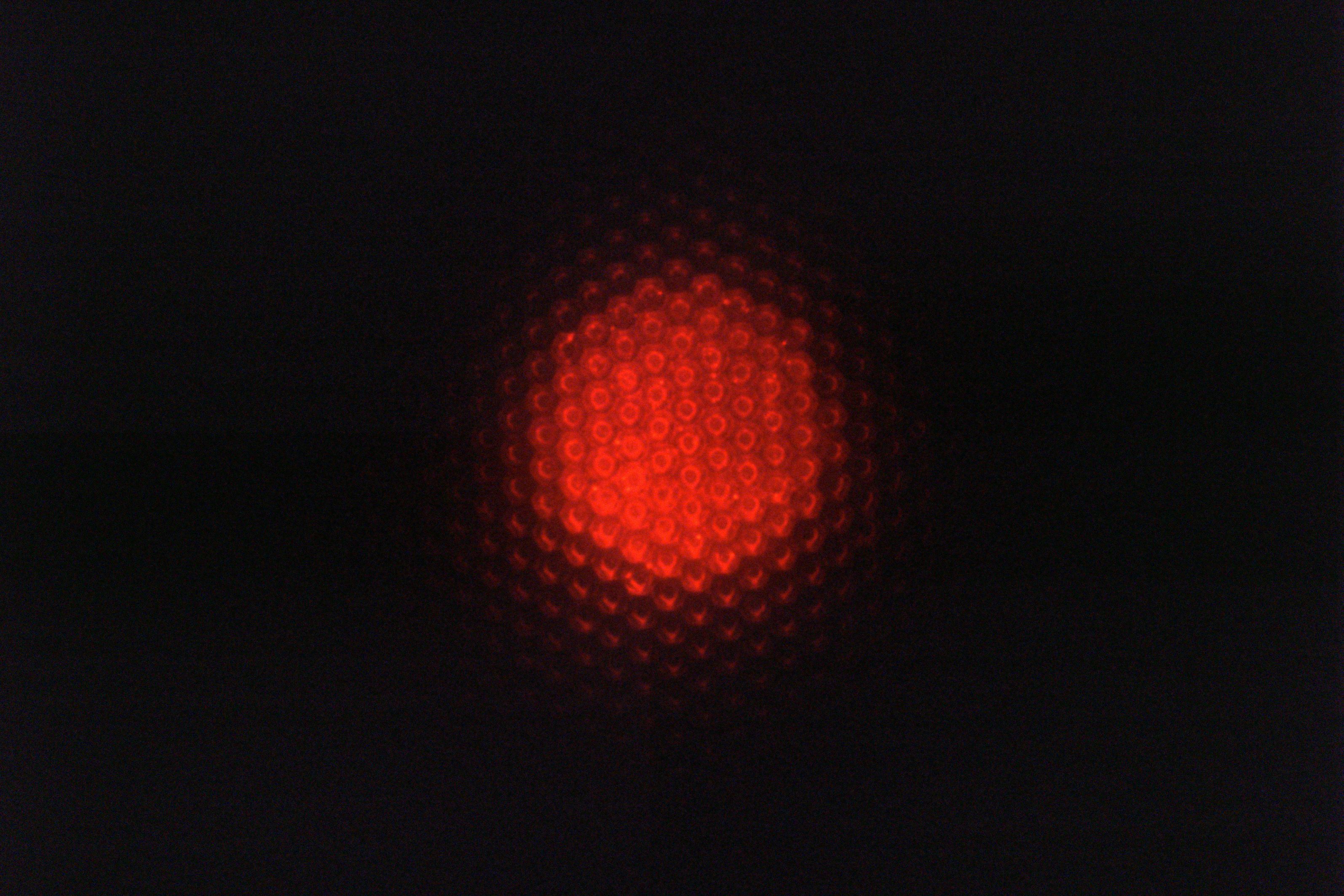
When Chazot had looked at the chip under a microscope, she noticed something curious: The chip emitted photons only at high angles forming a hollow cone of light. Turns out, the Bragg mirror had just the right layer thicknesses to only let photons pass through when they came at the mirror with a certain (high) angle.
“Once we saw this hollow cone of light, we wondered: ‘Could this device be useful for something?’” Chazot says. “And the answer was: Yes!”
As it turns out, they had incorporated the capabilities of multiple expensive, bulky dark-field microscope components into a single small chip.
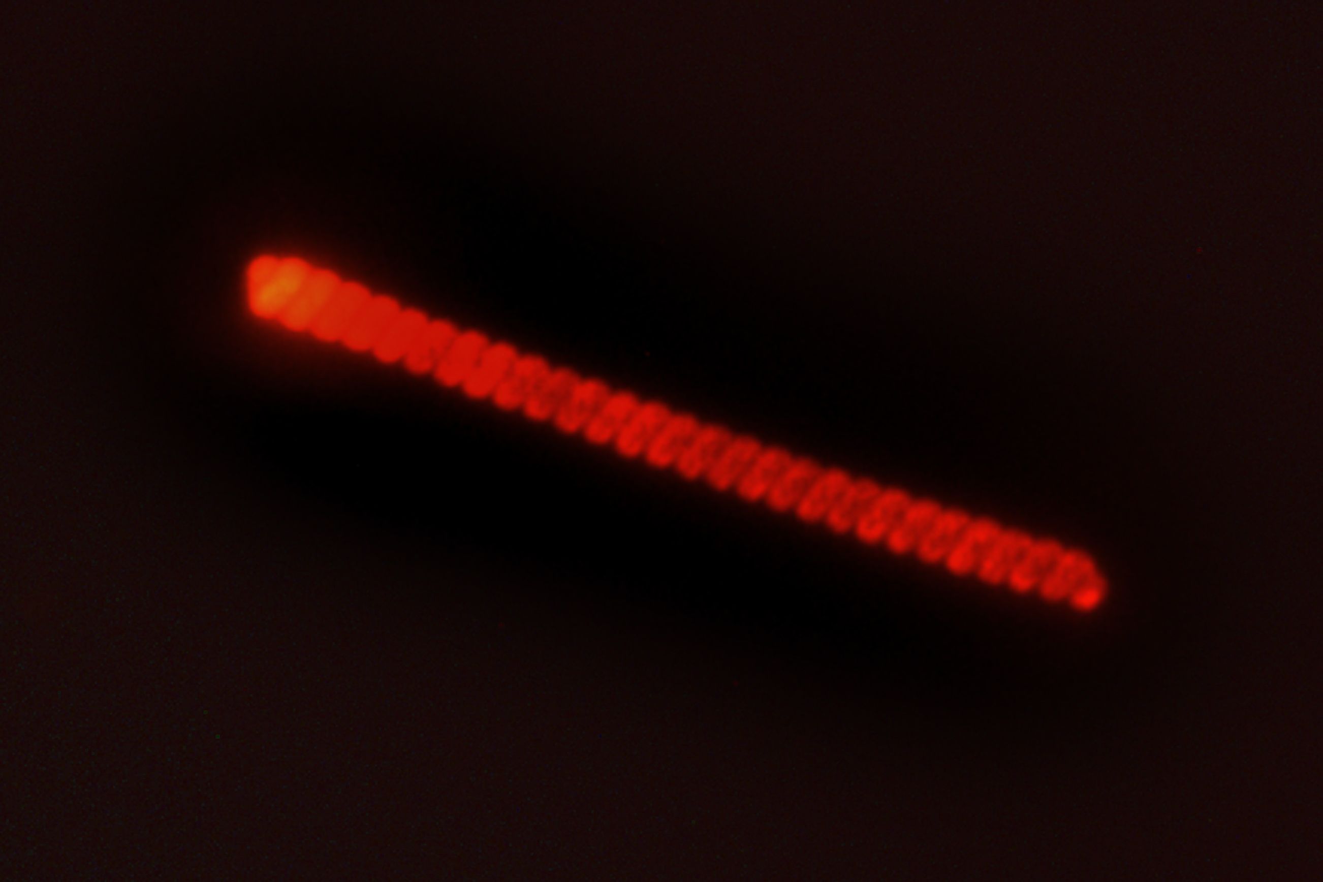
Chazot and her colleagues used well-established theoretical optical concepts to model the chip’s optical properties to optimize its performance for this newly found task. They fabricated multiple chips, each producing a hollow cone of light with a tailored angular profile.
“Regardless of the microscope you’re using, among all these tiny little chips, one will work with your objective,” Chazot says.
To test the chips, the team collected samples of seawater as well as nonpathogenic strains of the bacteria E. coli, and placed each sample on a chip that they set on the platform of a standard bright-field microscope. With this simple setup, they were able to produce clear and detailed dark-field images of individual bacterial cells, as well as microorganisms in seawater, which were close to invisible under bright-field illumination.
In the near future these dark-field illumination chips could be mass-produced and tailored for even simple, high school-grade microscopes, to enable imaging of low-contrast, translucent biological samples. In combination with other work in Kolle’s lab, the chips may also be incorporated into miniaturized dark-field imaging devices for point-of-care diagnostics and bioanalytical applications in the field.
“This is a wonderful story of discovery based innovation that has the potential for widespread impact in science and education through outfitting garden-variety microscopes with this technology,” says James Burgess, program manager for the Institute for Soldier Nanotechnologies, Army Research Office. “Additionally, the ability to obtain superior contrast in imaging of biological and inorganic materials under optical magnification could be incorporated into systems for identification of new biological threats and toxins in Army Medical Center laboratories and on the battlefield.”
This research was supported, in part, by the National Science Foundation, the U.S. Army Research Office, and the National Institutes of Health.


