Many types of cancer could be more easily treated if they were detected at an earlier stage. MIT researchers have now developed an imaging system, named “DOLPHIN,” which could enable them to find tiny tumors, as small as a couple of hundred cells, deep within the body.
In a new study, the researchers used their imaging system, which relies on near-infrared light, to track a 0.1-millimeter fluorescent probe through the digestive tract of a living mouse. They also showed that they can detect a signal to a tissue depth of 8 centimeters, far deeper than any existing biomedical optical imaging technique.
The researchers hope to adapt their imaging technology for early diagnosis of ovarian and other cancers that are currently difficult to detect until late stages.
“We want to be able to find cancer much earlier,” says Angela Belcher, the James Mason Crafts Professor of Biological Engineering and Materials Science at MIT and a member of the Koch Institute for Integrative Cancer Research, and the newly-appointed head of MIT’s Department of Biological Engineering. “Our goal is to find tiny tumors, and do so in a noninvasive way.”
Belcher is the senior author of the study, which appears in the March 7 issue of Scientific Reports. Xiangnan Dang, a former MIT postdoc, and Neelkanth Bardhan, a Mazumdar-Shaw International Oncology Fellow, are the lead authors of the study. Other authors include research scientists Jifa Qi and Ngozi Eze, former postdoc Li Gu, postdoc Ching-Wei Lin, graduate student Swati Kataria, and Paula Hammond, the David H. Koch Professor of Engineering, head of MIT’s Department of Chemical Engineering, and a member of the Koch Institute.
Deeper imaging
Existing methods for imaging tumors all have limitations that prevent them from being useful for early cancer diagnosis. Most have a tradeoff between resolution and depth of imaging, and none of the optical imaging techniques can image deeper than about 3 centimeters into tissue. Commonly used scans such as X-ray computed tomography (CT) and magnetic resonance imaging (MRI) can image through the whole body; however, they can’t reliably identify tumors until they reach about 1 centimeter in size.
Belcher’s lab set out to develop new optical methods for cancer imaging several years ago, when they joined the Koch Institute. They wanted to develop technology that could image very small groups of cells deep within tissue and do so without any kind of radioactive labeling.
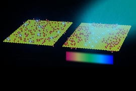
Near-infrared light, which has wavelengths from 900 to 1700 nanometers, is well-suited to tissue imaging because light with longer wavelengths doesn’t scatter as much as when it strikes objects, which allows the light to penetrate deeper into the tissue. To take advantage of this, the researchers used an approach known as hyperspectral imaging, which enables simultaneous imaging in multiple wavelengths of light.
The researchers tested their system with a variety of near-infrared fluorescent light-emitting probes, mainly sodium yttrium fluoride nanoparticles that have rare earth elements such as erbium, holmium, or praseodymium added through a process called doping. Depending on the choice of the doping element, each of these particles emits near-infrared fluorescent light of different wavelengths.
Using algorithms that they developed, the researchers can analyze the data from the hyperspectral scan to identify the sources of fluorescent light of different wavelengths, which allows them to determine the location of a particular probe. By further analyzing light from narrower wavelength bands within the entire near-IR spectrum, the researchers can also determine the depth at which a probe is located. The researchers call their system “DOLPHIN”, which stands for “Detection of Optically Luminescent Probes using Hyperspectral and diffuse Imaging in Near-infrared.”
To demonstrate the potential usefulness of this system, the researchers tracked a 0.1-millimeter-sized cluster of fluorescent nanoparticles that was swallowed and then traveled through the digestive tract of a living mouse. These probes could be modified so that they target and fluorescently label specific cancer cells.
“In terms of practical applications, this technique would allow us to non-invasively track a 0.1-millimeter-sized fluorescently-labeled tumor, which is a cluster of about a few hundred cells. To our knowledge, no one has been able to do this previously using optical imaging techniques,” Bardhan says.
Earlier detection
The researchers also demonstrated that they could inject fluorescent particles into the body of a mouse or a rat and then image through the entire animal, which requires imaging to a depth of about 4 centimeters, to determine where the particles ended up. And in tests with human tissue-mimics and animal tissue, they were able to locate the probes to a depth of up to 8 centimeters, depending on the type of tissue.
Guosong Hong, an assistant professor of materials science and engineering at Stanford University, described the new method as “game-changing.”
“This is really amazing work,” says Hong, who was not involved in the research. “For the first time, fluorescent imaging has approached the penetration depth of CT and MRI, while preserving its naturally high resolution, making it suitable to scan the entire human body.”
This kind of system could be used with any fluorescent probe that emits light in the near-infrared spectrum, including some that are already FDA-approved, the researchers say. The researchers are also working on adapting the imaging system so that it could reveal intrinsic differences in tissue contrast, including signatures of tumor cells, without any kind of fluorescent label.
In ongoing work, they are using a related version of this imaging system to try to detect ovarian tumors at an early stage. Ovarian cancer is usually diagnosed very late because there is no easy way to detect it when the tumors are still small.
“Ovarian cancer is a terrible disease, and it gets diagnosed so late because the symptoms are so nondescript,” Belcher says. “We want a way to follow recurrence of the tumors, and eventually a way to find and follow early tumors when they first go down the path to cancer or metastasis. This is one of the first steps along the way in terms of developing this technology.”
The researchers have also begun working on adapting this type of imaging to detect other types of cancer such as pancreatic cancer, brain cancer, and melanoma.
The research was funded by the Koch Institute Frontier Research Program, the Marble Center for Cancer Nanomedicine, the Koch Institute Support (core) Grant from the National Cancer Institute, the NCI Center for Center for Cancer Nanotechnology Excellence, and the Bridge Project.