A hundred years ago, “2d” meant a two-penny, or 1-inch, nail. Today, “2-D” encompasses a broad range of atomically thin flat materials, many with exotic properties not found in the bulk equivalents of the same materials, with graphene — the single-atom-thick form of carbon — perhaps the most prominent. While many researchers at MIT and elsewhere are exploring two-dimensional materials and their special properties, Frances M. Ross, the Ellen Swallow Richards Professor in Materials Science and Engineering, is interested in what happens when these 2-D materials and ordinary 3-D materials come together.
“We’re interested in the interface between a 2-D material and a 3-D material because every 2-D material that you want to use in an application, such as an electronic device, still has to talk to the outside world, which is three-dimensional,” Ross says.
“We’re at an interesting time because there are immense developments in instrumentation for electron microscopy, and there is great interest in materials with very precisely controlled structures and properties, and these two things cross in a fascinating way,” says Ross.
“The opportunities are very exciting,” Ross says. “We’re going to be really improving the characterization capabilities here at MIT.” Ross specializes in examining how nanoscale materials grow and react in both gases and liquid media, by recording movies using electron microscopy. Microscopy of reactions in liquids is particularly useful for understanding the mechanisms of electrochemical reactions that govern the performance of catalysts, batteries, fuel cells, and other important technologies. “In the case of liquid phase microscopy, you can also look at corrosion where things dissolve away, while in gases you can look at how individual crystals grow or how materials react with, say, oxygen,” she says.
Ross joined the Department of Materials Science and Engineering (DMSE) faculty last year, moving from the Nanoscale Materials Analysis department at the IBM Thomas J. Watson Research Center. “I learned a tremendous amount from my IBM colleagues and hope to extend our research in material design and growth in new directions,” she says.
Recording movies
During a recent visit to her lab, Ross explained an experimental setup donated to MIT by IBM. An ultra-high vacuum evaporator system arrived first, to be attached later directly onto a specially designed transmission electron microscope. “This gives powerful possibilities,” Ross explains. “We can put a sample in the vacuum, clean it, do all sorts of things to it such as heating and adding other materials, then transfer it under vacuum into the microscope, where we can do more experiments while we record images. So we can, for example, deposit silicon or germanium, or evaporate metals, while the sample is in the microscope and the electron beam is shining through it, and we are recording a movie of the process.”
While waiting this spring for the transmission electron microscope to be set up, members of Ross’ seven-member research group, including materials science and engineering postdoc Shu Fen Tan and graduate student Kate Reidy, made and studied a variety of self-assembled structures. The evaporator system was housed temporarily on the fifth-level prototyping space of MIT.nano while Ross’s lab was being readied in Building 13. “MIT.nano had the resources and space; we were happy to be able to help,” says Anna Osherov, MIT.nano assistant director of user services.
“All of us are interested in this grand challenge of materials science, which is: ‘How do you make a material with the properties you want and, in particular, how do you use nanoscale dimensions to tweak the properties, and create new properties, that you can’t get from bulk materials?’” Ross says.
Using the ultra-high vacuum system, graduate student Kate Reidy formed structures of gold and niobium on several 2-D materials. “Gold loves to grow into little triangles,” Ross notes. “We’ve been talking to people in physics and materials science about which combinations of materials are the most important to them in terms of controlling the structures and the interfaces between the components in order to give some improvement in the properties of the material,” she notes.
Shu Fen Tan synthesized nickel-platinum nanoparticles and examined them using another technique, liquid cell electron microscopy. She could arrange for only the nickel to dissolve, leaving behind spiky skeletons of platinum. “Inside the liquid cell, we are able to see this whole process at high spatial and temporal resolutions,” Tan says. She explains that platinum is a noble metal and less reactive than nickel, so under the right conditions the nickel participates in an electrochemical dissolution reaction and the platinum is left behind.
Platinum is a well-known catalyst in organic chemistry and fuel cell materials, Tan notes, but it is also expensive, so finding combinations with less-expensive materials such as nickel is desirable.
“This is an example of the range of materials reactions you can image in the electron microscope using the liquid cell technique,” Ross says. “You can grow materials; you can etch them away; you can look at, for example, bubble formation and fluid motion.”
A particularly important application of this technique is to study cycling of battery materials. “Obviously, I can’t put an AA battery in here, but you could set up the important materials inside this very small liquid cell and then you can cycle it back and forth and ask, if I charge and discharge it 10 times, what happens? It does not work just as well as before — how does it fail?” Ross asks. “Some kind of failure analysis and all the intermediate stages of charging and discharging can be observed in the liquid cell.”
“Microscopy experiments where you see every step of a reaction give you a much better chance of understanding what’s going on,” Ross says.
Moiré patterns
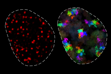
Graduate student Reidy is interested in how to control the growth of gold on 2-D materials such as graphene, tungsten diselenide, and molybdenum disulfide. When she deposited gold on “dirty” graphene, blobs of gold collected around the impurities. But when Reidy grew gold on graphene that had been heated and cleaned of impurities, she found perfect triangles of gold. Depositing gold on both the top and bottom sides of clean graphene, Reidy saw in the microscope features known as moiré patterns, which are caused when the overlapping crystal structures are out of alignment.
The gold triangles may be useful as photonic and plasmonic structures. “We think this could be important for a lot of applications, and it is always interesting for us to see what happens,” Reidy says. She is planning to extend her clean growth method to form 3-D metal crystals on stacked 2-D materials with various rotation angles and other mixed-layer structures. Reidy is interested in the properties of graphene and hexagonal boron nitride (hBN), as well as two materials that are semiconducting in their 2-D single-layer form, molybdenum disulfide (MoS2) and tungsten diselenide (WSe2). “One aspect that’s very interesting in the 2-D materials community is the contacts between 2-D materials and 3-D metals,” Reidy says. “If they want to make a semiconducting device or a device with graphene, the contact could be ohmic for the graphene case or a Schottky contact for the semiconducting case, and the interface between these materials is really, really important.”
“You can also imagine devices using the graphene just as a spacer layer between two other materials,” Ross adds.
For device makers, Reidy says it is sometimes important to have a 3-D material grow with its atomic arrangement aligned perfectly with the atomic arrangement in the 2-D layer beneath. This is called epitaxial growth. Describing an image of gold grown together with silver on graphene, Reidy explains, “We found that silver doesn’t grow epitaxially, it doesn’t make those perfect single crystals on graphene that we wanted to make, but by first depositing the gold and then depositing silver around it, we can almost force silver to go into an epitaxial shape because it wants to conform to what its gold neighbors are doing.”
Electron microscope images can also show imperfections in a crystal such as rippling or bending, Reidy notes. “One of the great things about electron microscopy is that it is very sensitive to changes in the arrangement of the atoms,” Ross says. “You could have a perfect crystal and it would all look the same shade of gray, but if you have a local change in the structure, even a subtle change, electron microscopy can pick it up. Even if the change is just within the top few layers of atoms without affecting the rest of the material beneath, the image will show distinctive features that allow us to work out what’s going on.”
Reidy also is exploring the possibilities of combining niobium — a metal that is superconducting at low temperatures — with a 2-D topological insulator, bismuth telluride. Topological insulators have fascinating properties whose discovery resulted in the Nobel Prize in Physics in 2016. “If you deposit niobium on top of bismuth telluride, with a very good interface, you can make superconducting junctions. We’ve been looking into niobium deposition, and rather than triangles we see structures that are more dendritic looking,” Reidy says. Dendritic structures look like the frost patterns formed on the inside of windows in winter, or the feathery patterns of some ferns. Changing the temperature and other conditions during the deposition of niobium can change the patterns that the material takes.
All the researchers are eager for new electron microscopes to arrive at MIT.nano to give further insights into the behavior of these materials. “Many things will happen within the next year, things are ramping up already, and I have great people to work with. One new microscope is being installed now in MIT.nano and another will arrive next year. The whole community will see the benefits of improved microscopy characterization capabilities here,” Ross says.
MIT.nano’s Osherov notes that two cryogenic transmission electron microscopes (cryo-TEM) are installed and running. “Our goal is to establish a unique microscopy-centered community. We encourage and hope to facilitate a cross-pollination between the cryo-EM researchers, primarily focused on biological applications and ‘soft’ material, as well as other research communities across campus,” she says. The latest addition of a scanning transmission electron microscope with enhanced analytical capabilities (ultrahigh energy resolution monochromator, 4-D STEM detector, Super-X EDS detector, tomography, and several in situ holders) brought in by John Chipman Associate Professor of Materials Science and Engineering James M. LeBeau, once installed, will substantially enhance the microscopy capabilities of the MIT campus. “We consider Professor Ross to be an immense resource for advising us in how to shape the in situ approach to measurements using the advanced instrumentation that will be shared and available to all the researchers within the MIT community and beyond,” Osherov says.
Little drinking straws
“Sometimes you know more or less what you are going to see during a growth experiment, but very often there’s something that you don’t expect,” Ross says. She shows an example of zinc oxide nanowires that were grown using a germanium catalyst. Some of the long crystals have a hole through their centers, creating structures which are like little drinking straws, circular outside but with a hexagonally shaped interior. “This is a single crystal of zinc oxide, and the interesting question for us is why do the experimental conditions create these facets inside, while the outside is smooth?” Ross asks. “Metal oxide nanostructures have so many different applications, and each new structure can show different properties. In particular, by going to the nanoscale you get access to a diverse set of properties.”
“Ultimately, we’d like to develop techniques for growing well-defined structures out of metal oxides, especially if we can control the composition at each location on the structure,” Ross says. A key to this approach is self-assembly, where the material builds itself into the structure you want without having to individually tweak each component. “Self-assembly works very well for certain materials but the problem is that there’s always some uncertainty, some randomness or fluctuations. There’s poor control over the exact structures that you get. So the idea is to try to understand self-assembly well enough to be able to control it and get the properties that you want,” Ross says.
“We have to understand how the atoms end up where they are, then use that self-assembly ability of atoms to make a structure we want. The way to understand how things self-assemble is to watch them do it, and that requires movies with high spatial resolution and good time resolution,” Ross explains. Electron microscopy can be used to acquire structural and compositional information and can even measure strain fields or electric and magnetic fields. “Imagine recording all of these things, but in a movie where you are also controlling how materials grow within the microscope. Once you have made a movie of something happening, you analyze all the steps of the growth process and use that to understand which physical principles were the key ones that determined how the structure nucleated and evolved and ended up the way it does.”
Future directions
Ross hopes to bring in a unique high-resolution, high vacuum TEM with capabilities to image materials growth and other dynamic processes. She intends to develop new capabilities for both water-based and gas-based environments. This custom microscope is still in the planning stages but will be situated in one of the rooms in the Imaging Suite in MIT.nano.
“Professor Ross is a pioneer in this field,” Osherov says. “The majority of TEM studies to-date have been static, rather than dynamic. With static measurements you are observing a sample at one particular snapshot in time, so you don’t gain any information about how it was formed. Using dynamic measurements, you can look at the atoms hopping from state to state until they find the final position. The ability to observe self-assembling processes and growth in real time provides valuable mechanistic insights. We’re looking forward to bringing these advanced capabilities to MIT.nano.” she says.
“Once a certain technique is disseminated to the public, it brings attention,” Osherov says. “When results are published, researchers expand their vision of experimental design based on available state-of-the-art capabilities, leading to many new experiments that will be focused on dynamic applications.”
Rooms in MIT.nano feature the quietest space on the MIT campus, designed to reduce vibrations and electromagnetic interference to as low a level as possible. “There is space available for Professor Ross to continue her research and to develop it further,” Osherov says. “The ability of in situ monitoring the formation of matter and interfaces will find applications in multiple fields across campus, and lead to a further push of the conventional electron microscopy limits.”