The following press release was issued today by the Broad Institute of MIT and Harvard.
Scientists routinely use genetic screens to perturb, or change the activity of, genes in mammalian cells, one at a time, to learn what those genes do. Pooled screens take this same approach but typically involve many more genetic perturbations across the whole genome. However, with pooled screens, scientists could only track cell survival and other simple whole-cell measurements.
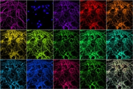
Microscopy is an important tool that allows biologists to simultaneously measure many cell features with high resolution. Researchers have sought to combine genetic screens with microscopy to study the effects of genes on a wide range of cellular behaviors, but doing this one gene at a time has been costly and time-consuming.
Now, researchers at the Broad Institute of MIT and Harvard and at MIT have developed a new method that combines large-scale pooled genetic screens with image-based analysis of cellular behavior. The approach, called optical pooled screens, allows researchers to examine how genes affect complex cellular processes with spatial and temporal resolution, in ways that other pooled screens could not.
“With this new approach, anyone can use microscopy to screen thousands of genes in a few days without specialized equipment,” said Avtar Singh, a postdoct in the lab of Paul Blainey at the Broad.
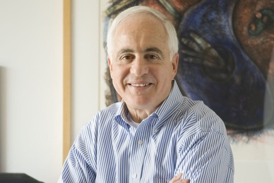
The study, published in Cell, was led by senior author Blainey, a core institute member at the Broad and associate professor in the Department of Biological Engineering at MIT. Singh was co-first author along with David Feldman, who was previously a physics PhD student in the labs of Feng Zhang and Blainey.
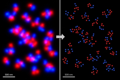
The authors examined the effects of 952 genes on the signaling activity of an immune-regulating complex called NF-kB by imaging the cellular location of a protein called p65 in millions of cells. They uncovered a new role for two genes, MED12 and MED24, in the relaxation of NFkB signaling.
“Looking through a microscope at live cells lets you see how genes influence biological processes in space and time — something you couldn't easily capture by other methods,” said Feldman.