It's a time of small marvels and big ideas. Welcome to the Nano Age.
MIT’s preparations for this new era are in full swing, including the recent launch of MIT.nano, the Institute's center for nanoscience and nanotechnology. And on the day after MIT.nano’s opening ceremonies, the Department of Biology hosted its Cryogenic Electron Microscopy (Cryo-EM) Symposium, which was co-organized by biology professor Thomas Schwartz and the director of the new facility, Edward Brignole.
“We organized the symposium to raise awareness of this new research capacity, and to celebrate the many people who worked to fund these instruments, design the space, build the suites, and set up the microscopes,” Brignole said of the Oct. 5 event. “We also wanted to bring together the various groups across MIT working on diverse technologies to improve Cryo-EM, from mathematicians, computer scientists, and electrical engineers to biologists, chemists, and biological engineers.”
The event featured pioneers leveraging Cryo-EM for various interdisciplinary applications both on campus and outside of MIT — from biology and machine learning to quantum mechanics.
The program included Ed Egelman from the University of Virginia, Mark Bathe from the MIT Department of Biological Engineering, Katya Heldwein from Tufts University's School of Medicine, and Karl Berggren from the Department of Electrical Engineering and Computer Science. Also giving talks were computational and systems biology graduate student Tristan Bepler from MIT's Computer Science and Artificial Intelligence Laboratory, Luke Chao from Harvard Medical School and Massachusetts General Hospital, postdoc Kuang Shen from the Whitehead Institute at MIT, and graduate student Jonathan Weed from the MIT Department of Mathematics. The talks were followed by a reception in Building 68 and guided tours of the Cryo-EM Facility.
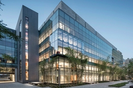
Unlike other popular techniques for determining 3-D macromolecular structures, Cryo-EM permits researchers to visualize more diverse and complex molecular assemblies in multiple conformations. Cryo-EM is housed in precisely climate-controlled rooms in the basement of MIT.nano, built atop a 5 million pound slab of concrete to minimize vibrations. Two multimillion-dollar instruments are being installed that will enable scientists to analyze cellular machinery in near-atomic detail; the microscopes are the first instruments to be installed in MIT.nano.
As Schwartz explained to an audience of more than 100 people during his opening remarks, he and his colleagues realized they needed to bring this technology to the MIT community. Like many of the center’s other tools, they would be too costly to purchase and too onerous to maintain for a single researcher or lab.
“Microscopes are very special and expensive tools, so this endeavor turned out to be much more involved than anything else I have done during my 14 years at MIT,” he said. “But this was not an effort of one or two people, it really took a whole community. We have many people to thank today.”
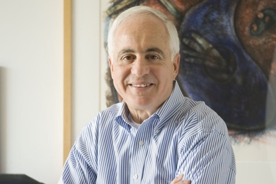
Establishing the Cryogenic Electron Microscopy Facility at MIT has been a long-time dream for Catherine Drennan, a professor of chemistry and biology and a Howard Hughes Medical Institute investigator. At the symposium, Drennan spoke about her work using the microscopes to capture snapshots of enzymes in action.
She remembers it was a “happy coincidence” that the plans for MIT.nano and the Cryo-EM Facility unfolded around the same time and then merged together to become one multi-disciplinary effort. Drennan, Schwartz, and others worked closely with MIT.nano Founding Director Vladimir Bulović and Vice President for Research Maria Zuber to gain institutional support and jumpstart the project. It took six years to design and construct MIT.nano, and four to implement the Cryo-EM Facility.
“We had this vision that the Cryo-EM Facility would be a shared space that people from all around MIT would use,” Drennan said.
An anonymous donor offered $5 million to fund the first microscope, the Titan Krios, while the Arnold and Mabel Beckman Foundation contributed $2.5 million to purchase the second, the Talos Arctica.
“The Beckman Foundation is really pleased to be supporting this kind of technology,” said Anne Hultgren, the foundation's executive director, who attended the symposium. “It was a win-win in terms of the timing and the need in the community. We are excited to be part of this effort, and to drive forward new innovations and experiments.”
The Beckman Foundation has made similar instrumentation grants to Johns Hopkins University School of Medicine, University of Pennsylvania’s Perelman School of Medicine, the University of Utah, and the University of Washington School of Medicine.
Drennan said that as the revolution in resolution continues to build, she hopes MIT’s new microscopes will bolster the resurging Cryo-EM community that’s slowly growing in and around Boston.
“Thanks to facilities like this, the Boston area went from being way behind the curve to right in front of it,” she said. “It's an incredibly exciting time, and I can’t wait to see how we learn and grow as a research community.”