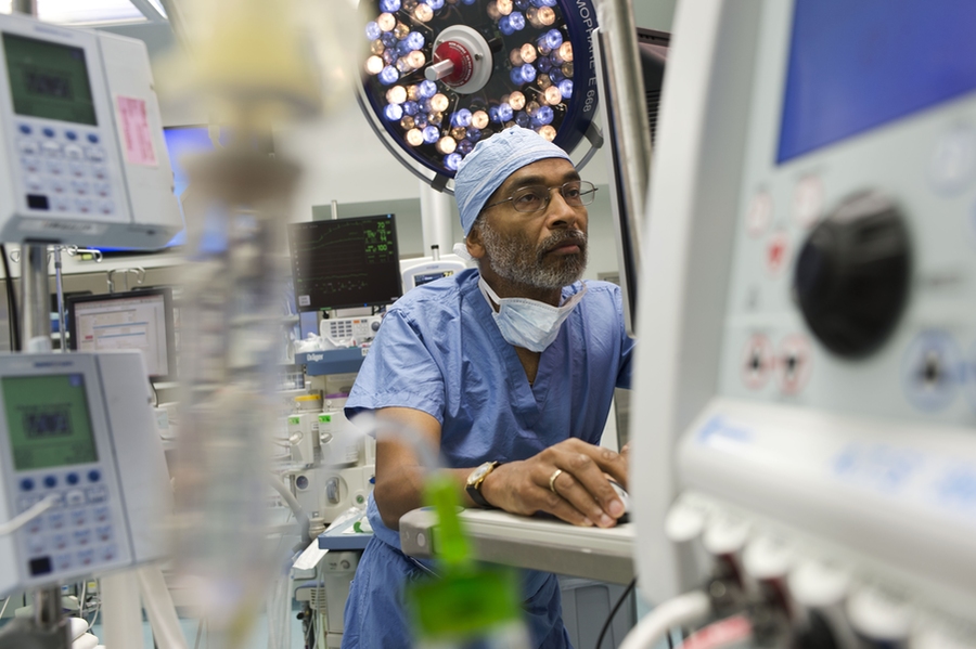
It’s intuitive that anesthesia operates in the brain, but the standard protocol among anesthesiologists when monitoring and dosing patients during surgery is to rely on indirect signs of arousal such as movement, and changes in heart rate and blood pressure. Through research in brain science and statistical modeling, Emery N. Brown, an anesthesiologist at Massachusetts General Hospital and neuroscientist at MIT’s Picower Institute for Learning and Memory, is putting the brain at the center of the field.
His findings allow him to safely give less anesthesia, for example, which can have important benefits for patients.
The key has been to develop a theoretical (i.e. neuroscientific) and analytical (i.e. statistical) understanding of electroencephalogram (EEG) brain wave measurements of patients under general anesthesia. In a presentation at the annual meeting of the American Association for the Advancement of Science in Austin, Texas, on Feb. 16, Brown described how anesthesia’s effects in the brain produce specific patterns of brain waves and how monitoring them via EEG data can lead to better care. He spoke as part of a broader discussion on the use of data analysis in brain research.
“We should use neuroscience and neuroscience paradigms to try to understand what’s happening in the brain under general anesthesia,” said Brown, the Edward Hood Taplin Professor of Medical Engineering and Computational Neuroscience in the Institute for Medical Engineering and Science and the Department of Brain and Cognitive Sciences at MIT. “It’s a neurophysiological process that affects the brain and central nervous system, so how can it be that what’s being developed in the neuroscience field is not being brought to bear to the question of the brain under anesthesia?”
In numerous papers over more than a decade, Brown has helped the field understand how different anesthesia drugs, such as propofol, dexmedetomidine, and sevofluran, interact with various neuronal receptors, affecting circuits in different regions of the brain. Those neurophysiological effects ultimately give rise to a state of unconsciousness — essentially a “reversible coma” — characterized by powerful, low frequency brain waves that essentially overwhelm the normal rhythms that synchronize various brain functions including sensory perception, higher cognition, and motor control.
Understanding anesthesia to this degree allows for practical insights. In an October 2016 study in in the Proceedings of the National Academy of Sciences, for example, Brown and colleague Ken Solt showed how stimulating dopamine-producing neurons in the ventral tegmental area of the brain could wake mice up from general anesthesia. The study suggests a way human patients could be awakened as well, which could lessen side effects, recover normal brain function more rapidly and help patients move more quickly out of the operating room and into recovery.
In parallel with illuminating the neuroscience of general anesthesia, Brown has developed statistical methods to finely analyze EEG measurements to the point where anesthesiologists can apply that knowledge to patients. Brown has shown, for instance, that EEG readings of level of unconsciousness vary in characteristic ways based on the drug, its dose, and the patient’s age.
“The deciphering of how these drugs are acting in the brain turns out to be an important signal processing question,” Brown said. “The drugs work by producing oscillations, these oscillations are readily visible in the EEG and they change very systematically with drug dose, class, and age.”
During the course of every surgery, Brown said, he uses real-time EEG readings to keep a patient adequately dosed without giving too much. In a recent case involving an 81-year-old cancer patient, Brown said he was able to comfortably administer about a third the supposedly needed dose. This can be especially important for older patients.
“We already know you don’t have to give older people as much, but it turns out it can be even less,” said Brown, who is one of the founding investigators of MIT’s Aging Brain Initiative.
Older patients are especially susceptible to problematic side effects when they wake up including delirium or post-operative cognitive dysfunction. Neuroscientifically informed ways to prevent giving too much anesthesia can help prevent such problems, Brown said.
In his latest paper, published last month in the Proceedings of the National Academy of Sciences , Brown’s group led by postdoctoral fellow Seong-Eun Kim presented a powerful new algorithm for analyzing data sets, like EEGs, where waves vary over time. The SS-MT method produces high-resolution, low-noise spectrograms from such data. In the study, he used SS-MT to discern variations in EEGs with different states of consciousness in patients who had received propofol.
“If we can get clearer, cleaner noise-free measures of the spectrogram then we can infer the states even better,” he said.
Brown said he doesn’t want his methods to stay just with him. To help put the brain at the center of practice, Brown has developed training materials and made them freely available at http://www.anesthesiaeeg.com. He is also working to advance these ideas within professional societies.
Ultimately, as more anesthesiologists acquire knowledge and EEG equipment, and equipment makers produce better displays for their use, Brown said, the field can move to a model where doctors have a direct view of the patient’s brain when monitoring and maintaining their consciousness during surgery.