
Every year 40,000 women die from breast cancer in the U.S. alone. When cancers are found early, they can often be cured. Mammograms are the best test available, but they’re still imperfect and often result in false positive results that can lead to unnecessary biopsies and surgeries.
One common cause of false positives are so-called “high-risk” lesions that appear suspicious on mammograms and have abnormal cells when tested by needle biopsy. In this case, the patient typically undergoes surgery to have the lesion removed; however, the lesions turn out to be benign at surgery 90 percent of the time. This means that every year thousands of women go through painful, expensive, scar-inducing surgeries that weren’t even necessary.
How, then, can unnecessary surgeries be eliminated while still maintaining the important role of mammography in cancer detection? Researchers at MIT’s Computer Science and Artificial Intelligence Laboratory (CSAIL), Massachusetts General Hospital, and Harvard Medical School believe that the answer is to turn to artificial intelligence (AI).
As a first project to apply AI to improving detection and diagnosis, the teams collaborated to develop an AI system that uses machine learning to predict if a high-risk lesion identified on needle biopsy after a mammogram will upgrade to cancer at surgery.
When tested on 335 high-risk lesions, the model correctly diagnosed 97 percent of the breast cancers as malignant and reduced the number of benign surgeries by more than 30 percent compared to existing approaches.
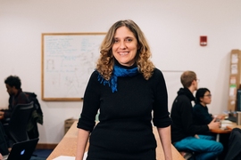
“Because diagnostic tools are so inexact, there is an understandable tendency for doctors to over-screen for breast cancer,” says Regina Barzilay, MIT’s Delta Electronics Professor of Electrical Engineering and Computer Science and a breast cancer survivor herself. “When there’s this much uncertainty in data, machine learning is exactly the tool that we need to improve detection and prevent over-treatment.”
Trained on information about more than 600 existing high-risk lesions, the model looks for patterns among many different data elements that include demographics, family history, past biopsies, and pathology reports.
“To our knowledge, this is the first study to apply machine learning to the task of distinguishing high-risk lesions that need surgery from those that don’t,” says collaborator Constance Lehman, professor at Harvard Medical School and chief of the Breast Imaging Division at MGH’s Department of Radiology. “We believe this could support women to make more informed decisions about their treatment, and that we could provide more targeted approaches to health care in general.”
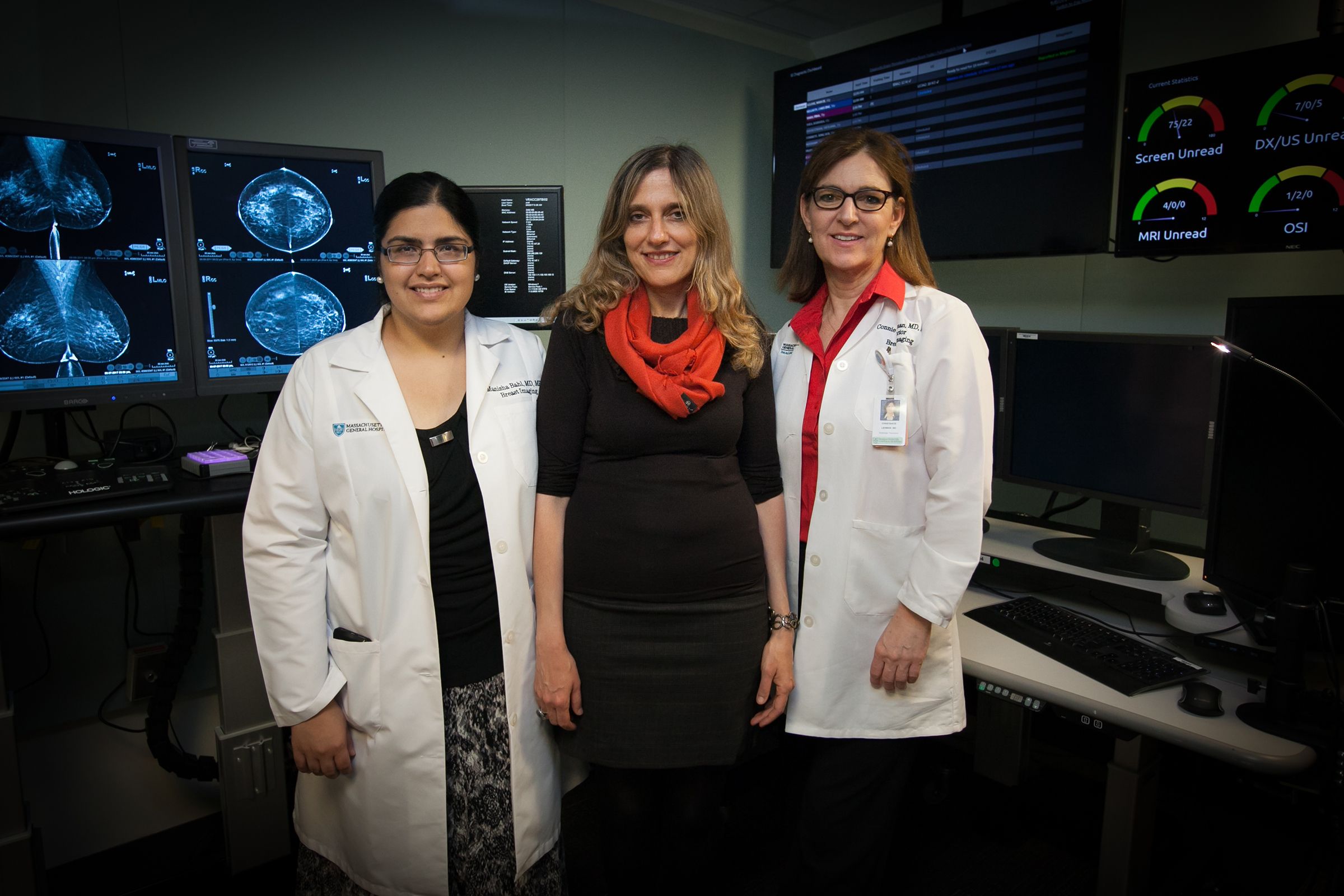
A recent MacArthur “genius grant” recipient, Barzilay is a co-author of a new journal article describing the results, co-written with Lehman and Manisha Bahl of MGH, as well as CSAIL graduate students Nicholas Locascio, Adam Yedidia, and Lili Yu. The article was published today in the medical journal Radiology.
How it works
When a mammogram detects a suspicious lesion, a needle biopsy is performed to determine if it is cancer. Roughly 70 percent of the lesions are benign, 20 percent are malignant, and 10 percent are high-risk lesions.
Doctors manage high-risk lesions in different ways. Some do surgery in all cases, while others perform surgery only for lesions that have higher cancer rates, such as “atypical ductal hyperplasia” (ADH) or a “lobular carcinoma in situ” (LCIS).
The first approach requires that the patient undergo a painful, time-consuming, and expensive surgery that is usually unnecessary; the second approach is imprecise and could result in missing cancers in high-risk lesions other than ADH and LCIS.
“The vast majority of patients with high-risk lesions do not have cancer, and we’re trying to find the few that do,” says Bahl, a fellow doctor at MGH’s Department of Radiology. “In a scenario like this there’s always a risk that when you try to increase the number of cancers you can identify, you’ll also increase the number of false positives you find.”
Using a method known as a “random-forest classifier,” the team's model resulted in fewer unnecessary surgeries compared to the strategy of always doing surgery, while also being able to diagnose more cancerous lesions than the strategy of only doing surgery on traditional “high-risk lesions.” (Specifically, the new model diagnosed 97 percent of cancers compared to 79 percent.)
“This work highlights an example of using cutting-edge machine learning technology to avoid unnecessary surgery,” says Marc Kohli, director of clinical informatics in the Department of Radiology and Biomedical Imaging at the University of California at San Francisco. “This is the first step toward the medical community embracing machine learning as a way to identify patterns and trends that are otherwise invisible to humans.”
Lehman says that MGH radiologists will begin incorporating the model into their clinical practice over the next year.
“In the past we might have recommended that all high-risk lesions be surgically excised,” Lehman says. “But now, if the model determines that the lesion has a very low chance of being cancerous in a specific patient, we can have a more informed discussion with our patient about her options. It may be reasonable for some patients to have their lesions followed with imaging rather than surgically excised.”
The team says that they are still working to further hone the model.
“In future work we hope to incorporate the actual images from the mammograms and images of the pathology slides, as well as more extensive patient information from medical records,” says Bahl.
Moving forward, the model could also easily be tweaked to be applied to other kinds of cancer and even other diseases entirely.
“A model like this will work anytime you have lots of different factors that correlate with a specific outcome,” says Barzilay. “It hopefully will enable us to start to go beyond a one-size-fits-all approach to medical diagnosis.”