What do engineers see when they look at their nanocrystal, microneedle, and quantum-dot experiments? Results, of course.
In a June workshop administered by the Department of Chemical Engineering, a group of graduate students worked with Felice Frankel, a research scientist at the Center for Materials Science and Engineering and an award-winning photographer, to improve both their technical skills and their aesthetic sensibilities and to create engaging and appealing photos of their work. Drawing from the fields of chemistry, materials science, and chemical engineering, the graduate-level researchers created powerful visuals that are intimately linked to the work that motivates and compels them.
Here are a few examples of their images.
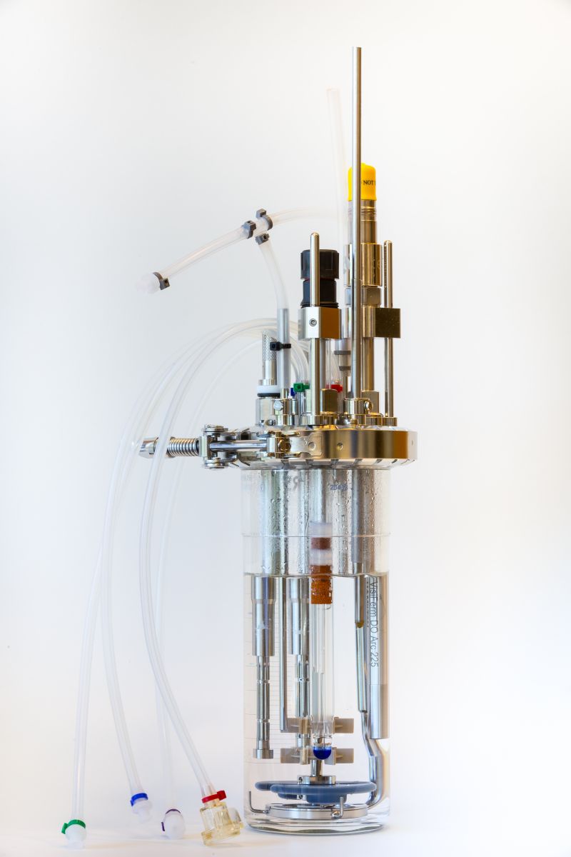
"This is a fully outfitted bioreactor designed for the production of protein-based therapeutics. This reactor enables the rapid manufacturing of multiple high-quality biologics.
As a graduate student, I’m interested in the modeling, simulation, optimization, and control of such systems, and this object is one of the more beautiful pieces of process equipment in the unit.
I learned a great deal about the use of light. In this case, diffuse light and the use of a white background gave the subject an even cast. I also enjoyed the discussion of ethical considerations of digital manipulation in photography, particularly with research photographs."
—Amos Enshen Lu, PhD candidate in chemical engineering

"These are semiconductor nanocrystals — also called quantum dots — suspended as a colloid in a solvent. Our group studies how these nanocrystals assemble when the solvent evaporates and how energy is transferred between them.
In this image, we excited the nanocrystals with an ultraviolet lamp. They absorb the high-energy ultraviolet light then emit light of their own, the color of which is a reflection of the nanocrystal size.
I learned how to photograph remissive samples like this, how to properly frame the image and background, and how to better accentuate the vial caps by using a directed white light to provide additional illumination from above."
—Aaron Goodman, graduate student in chemistry, and Mark Weidman, graduate student in chemical engineering

"This is a stretchable fluidic device imprinted in a tough hydrogel and closed on top with a thin elastomer layer. Similar devices can be used as platforms for biological and tissue engineering experiments by changing the designs patterned on the hydrogel.
The angle and framing make this image better than any I’ve done before. I learned about the different options for composing my image, and that changing the background and lighting can make drastic differences in the final look of the device. By making the right choices, a photo of a standard research device can become a cover page."
—German Alberto Parada Hernandez, graduate student in chemical engineering

"These are microneedles, which are mostly used for the delivery of drugs and vaccines across the skin, and the silicone mold used to make them. Microneedles provide greater insight into the monitoring of immunity and can aid in the design of new vaccines. I am using them in my research to monitor the immune system and for other disease-related diagnostics.
I experimented with different lighting conditions, backgrounds, and angles to get an engaging and crisp image of my scientific endeavors."
—Anasuya Mandal, graduate student in chemical engineering

"These are microscope slides coated with hundreds of polymer patches containing catalase, an antioxidant enzyme, soaking in a solution of hydrogen peroxide. The bubbles form when catalase converts the hydrogen peroxide into water and oxygen. These polymer patches can be attached to the surface of living cells and carried throughout the body as 'cell backpacks.'
Our goal is to explore the use of these backpacks for cell-mediated and targeted drug delivery. They enable the non-invasive delivery of drugs to sites of inflammation, even across the blood-brain barrier, which is a significant obstacle to traditional drug treatment.
I learned how to create a composition and use visuals to illustrate scientific concepts — and, in turn, share my research work with a much larger audience."
—Roberta Polak, postdoc in materials science and engineering

"This flow device represents a crucial step in the scale-up of the electrochemical separation processes developed by the Hatton group. The electrochemical cell contains a sandwich redox-electrode configuration, and serves to validate our water purification and remediation technology under flow conditions.
We hope to address challenges such as the removal of emerging contaminants in pesticides, pharmaceuticals, and heavy-metal micropollutants.
By experimenting with visuals, I learned how to better communicate my work. By optimizing camera settings, lighting conditions, and composition, I can now produce an appealing final product to a wider audience."
—Xiao Su, graduate student in chemical engineering