Cells for our skin, hair, or nails share DNA that is the same or nearly identical, yet slight differences in which genes are expressed from that DNA give each cell its unique characteristics. MIT assistant professor of physics Ibrahim Cissé is studying how a specialized enzyme called RNA polymerase II controls this gene expression.
"All the DNA presumably are the same or almost identical. It is just the set of active genes, the set of messages, that make up a particular cell's identity," Cissé explains.
In normal cells, enzymes called polymerase do the work of transcription — that is, copying a gene from the DNA, and producing a messenger RNA molecule that is primed to make a protein according to the formula encoded in the gene.
His recent research suggests that short-lived clusters of RNA polymerase II that target a specific gene control how messenger RNA molecules get made.
Primary motivation
"First and foremost, we are driven by the idea of fundamentally understanding nature and how living matter works. That's what motivates us," Cissé says.
Cissé uses highly sensitive microscopy methods that look directly inside the nucleus of a living cell to show the behavior of single biomolecules. "What interests us are a class of biomolecular interactions that tend to be very weak; indeed they are called weak and transient interactions," he says.
"The more detail we have about biomolecular interactions, the more we can parse out what is special about them, and their function. We can understand how they actually work, rather than taking wild guesses about what they might be doing just because of some sort of average behavior that they exhibit," Cissé adds.
The research combines super-resolution microscopy, modifying genes to make proteins that are fluorescent, as well as computational techniques for maximizing image quality and analysis. "Everything that we've been doing in our lab, for the past year and a half now, has been almost exclusively in living cells, with this kind of single molecule super-resolution sensitivity," he says.
Crowd control
Cissé's research focuses on how RNA polymerase II (Pol II) functions. A single polymerase molecule does not do the work on its own but rather a polymerase molecule is assisted by other transcription factors that are all recruited at a DNA site to form what is called a "pre-initiation complex." The proper formation of this "big machine," as Cissé calls it, is necessary for a single polymerase molecule to bind to DNA and begin its work of copying or transcribing a specific gene segment.
"When the polymerase gets recruited, what it does is, through the help of the other factors, is form this complex that tracks on the DNA like a train on the railroad, and then makes a copy of just one of the strands of that (DNA). The RNA is a letter code copy just spelling out this portion of the DNA, which is the gene," he explains.
"There has been this fundamental question in the field, on how it is that a cell can receive an external signal and integrate that external signal through all these complex interactions, and determine that precisely this gene should be turned on. So how do you gain precision and control out of this very dynamic process? Now, we suspect that this transient Pol II crowding — clustering — that we uncovered could be one of those hidden knobs that the cell can use to tune gene expression," Cissé says.
A scientific paper on these findings is currently under review for publication. "We knew that cells could receive an external signal and generate this burst of gene transcripts. There was a missing link in how this control happened precisely at the gene level. And we suspect to have now found previously hidden clues in this process," Cissé says.
Extracting principles
In abnormally behaving cells, such as cancer cells, "the set of messages that the cell should be expressing is disregulated. The cell is no longer able to control precisely what genes are expressed and what those expressed genes actually do to the cell," Cissé explains.
"By being able to understand how gene expression actually happens — for the first time at this level of molecular details directly inside the living cell — if we understand how it happens normally, then maybe we can start understanding how it happens when things go wrong," Cissé continues. "We should hope to be able to finally unveil the slight details that make up the difference between a normal cell and a malignant cell."
"What we suspect is that the guiding principles we would unveil about how these low affinity interactions control gene expression, in this particular context, might probably lead to a more general understanding," Cissé says. "We aim to uncover general physical principles that govern how low affinity interactions work, what other, similar processes might be happening in the living cells, and how the disregulation of these processes may probably result in diseases."
Weak and transient interactions
A 2013 paper in Science, of which Cissé was lead author, showed RNA polymerase II clusters had an average lifetime of about 5 seconds. The study analyzed bone cancer cells using a technique called photoactivation localization microscopy (PALM).
"These weak interactions inside the living cell tend to control so-called genome regulation in ways we didn't even expect," Cissé explains. Protein clustering is a prominent feature in a wide range of cellular processes, and protein aggregates have been reported in poorly understood neurodegenerative diseases such as amyotrophic lateral sclerosis (ALS) and Alzheimer's disease; but studying them quantitatively in living cells has not been easy due to limitations in current techniques. "With our approaches, even if protein crowding is very fast and very dynamic, we can capture and study that now in living cells," Cissé says.
Super-resolution techniques
Cissé joined the MIT physics faculty in January 2014. A native of Niger, he received his BS in physics in 2004 from North Carolina Central University and his doctorate in physics from the University of Illinois at Urbana-Champaign in 2009. He was a fellow at ENS Paris from January 2010 to December 2012 and worked at the Transcription Imaging Consortium at Howard Hughes Medical Institute's Janelia Farm Research Campus in Ashburn, Virginia, in 2013.
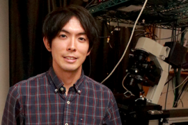
At MIT, Cissé has assembled an interdisciplinary team of postdocs and graduate students to pursue his research. Building on super-resolution microscope techniques that won the 2014 Nobel Prize in chemistry for Eric Betzig, Stefan W. Hell, and William E. Moerner, Cissé is seeking to push the method further. Graduate student Takuma Inoue helped build the laser bench setup for super-resolution microscopy in Cissé's lab. The researchers genetically modified the polymerase II enzyme to include a fluorescent marker that lets them image the polymerase. One color of laser light activates the fluorescent marker, known as a fluorophore, which then emits light of a different color allowing the researchers to track it. The group uses a derivative of the green fluorescent protein, a substance originally isolated from jellyfish. "When you shine one color of light on it, it glows in a different color. And since you aren't supplying anything with that color, you know that wherever you see this newly created light coming from, that has to be the location of one of these molecules," graduate student Owen Andrews says.
The lab uses lasers emitting light at 405 nanometers, which is a pale purple; 488 nanometers, a cyan blue; and 561 nanometers, a yellowish green. "We will take DNA for the different genes that we're interested in, and we'll modify them by attaching the sequence that codes for these fluorophores," Andrews explains.
The super-resolution imaging setup captures images through the microscope onto an electron-multiplying charge-coupled device (EM-CCD). "It can detect very sensitive signals, even single photons, and also it's a very fast camera," explains Inoue, who built the setup. The EM-CCD can acquire tens of thousands of images, each with a few millisecond exposure time, but overall it typically takes about eight minutes to get one super-resolution image.
Andrews is developing data analysis tools to extract more dynamic information from this data to precisely localize and processes much faster than the time it typically takes to get a super-resolution image. "Can we push both the technique and the analysis from the technique to gain more insight about these highly dynamic biological processes?" Cissé asks. "In terms of super-resolution, what was done previously with this technique, I would say, has been to image mostly static structures, for example in dead cells Our whole point now is going directly inside living cell and looking at those dynamics."
"We really have many different people who are working to address outstanding questions," Cissé adds. "There are existing techniques that we can use and improve upon to get us this far; but we know it will only get us this far. And then there are people in the lab, on the other hand, who are thinking about what we can do differently to go beyond what the best existing techniques allow us to do. I think that's actually what makes this group so exciting for me. There is no prescribed way to solve these challenging questions that interest us. But in the weekly group meetings there are almost always interesting new updates to discuss. We're continually thinking about where can we take this research, and what is maybe the best impact that this research can have," he says. Inoue, for example, also is working on new ways of tagging the RNA itself, without genetic modification, to get better images of it.
Pursuing what you're most excited about
Cissé's application of single-molecule techniques to study polymerase enzymes and gene expression is funded under the National Institutes of Health Project No. 1DP2CA195769-01, with additional funds from the National Cancer Institute.
"What's great about [the NIH Director's New Innovator Award] is it really gives you the flexibility of pursuing whatever you are most excited about. That award, for instance, provided the seed funding for the exploratory research that Takuma started," Cissé explains. The Department of Physics continues to fund the research.
"The obvious next step will be to figure out the details of how this polymerase clustering forms and how, maybe from that knowledge, we can think about possible ways of intervening. These could very well be the basis for possible therapies, and reveal new principles behind the regulation and control of gene expression in living cells," he suggests.
"Now, at best, we can look at the polymerase in one color and the RNA coming out in a different color," he says. But in a cell, when polymerase gets recruited, it is helped by dozens of other transcription factors. "If we can look at the polymerase and are seeing just part of the story, can we now try to understand the entire pre-initiation complex assembly?" Cissé asks. The lab is trying to extend this work through multi-color microscopy to analyze the polymerase clusters and related transcription factors in more detail.


