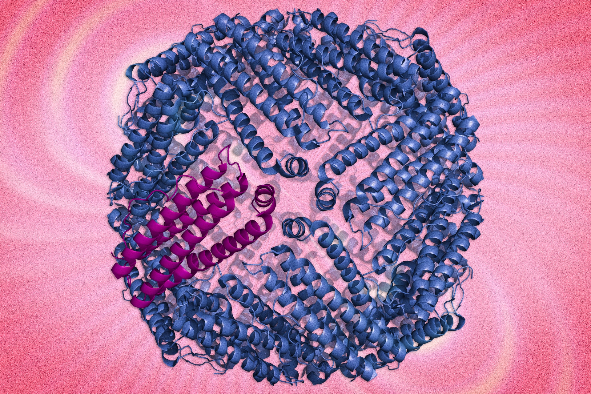

MIT engineers have designed magnetic protein nanoparticles that can be used to track cells or to monitor interactions within cells. The particles, described today in Nature Communications, are an enhanced version of a naturally occurring, weakly magnetic protein called ferritin.
“Ferritin, which is as close as biology has given us to a naturally magnetic protein nanoparticle, is really not that magnetic. That’s what this paper is addressing,” says Alan Jasanoff, an MIT professor of biological engineering and the paper’s senior author. “We used the tools of protein engineering to try to boost the magnetic characteristics of this protein.”
The new “hypermagnetic” protein nanoparticles can be produced within cells, allowing the cells to be imaged or sorted using magnetic techniques. This eliminates the need to tag cells with synthetic particles and allows the particles to sense other molecules inside cells.
The paper’s lead author is former MIT graduate student Yuri Matsumoto. Other authors are graduate student Ritchie Chen and Polina Anikeeva, an assistant professor of materials science and engineering.
Magnetic pull
Previous research has yielded synthetic magnetic particles for imaging or tracking cells, but it can be difficult to deliver these particles into the target cells.
In the new study, Jasanoff and colleagues set out to create magnetic particles that are genetically encoded. With this approach, the researchers deliver a gene for a magnetic protein into the target cells, prompting them to start producing the protein on their own.
“Rather than actually making a nanoparticle in the lab and attaching it to cells or injecting it into cells, all we have to do is introduce a gene that encodes this protein,” says Jasanoff, who is also an associate member of MIT’s McGovern Institute for Brain Research.
As a starting point, the researchers used ferritin, which carries a supply of iron atoms that every cell needs as components of metabolic enzymes. In hopes of creating a more magnetic version of ferritin, the researchers created about 10 million variants and tested them in yeast cells.
After repeated rounds of screening, the researchers used one of the most promising candidates to create a magnetic sensor consisting of enhanced ferritin modified with a protein tag that binds with another protein called streptavidin. This allowed them to detect whether streptavidin was present in yeast cells; however, this approach could also be tailored to target other interactions.
The mutated protein appears to successfully overcome one of the key shortcomings of natural ferritin, which is that it is difficult to load with iron, says Alan Koretsky, a senior investigator at the National Institute of Neurological Disorders and Stroke.
“To be able to make more magnetic indicators for MRI would be fabulous, and this is an important step toward making that type of indicator more robust,” says Koretsky, who was not part of the research team.
Sensing cell signals
Because the engineered ferritins are genetically encoded, they can be manufactured within cells that are programmed to make them respond only under certain circumstances, such as when the cell receives some kind of external signal, when it divides, or when it differentiates into another type of cell. Researchers could track this activity using magnetic resonance imaging (MRI), potentially allowing them to observe communication between neurons, activation of immune cells, or stem cell differentiation, among other phenomena.
Such sensors could also be used to monitor the effectiveness of stem cell therapies, Jasanoff says.
“As stem cell therapies are developed, it’s going to be necessary to have noninvasive tools that enable you to measure them,” he says. Without this kind of monitoring, it would be difficult to determine what effect the treatment is having, or why it might not be working.
The researchers are now working on adapting the magnetic sensors to work in mammalian cells. They are also trying to make the engineered ferritin even more strongly magnetic.