
About 10 percent of the U.S. population suffers from dyslexia, a condition that makes learning to read difficult. Dyslexia is usually diagnosed around second grade, but the results of a new study from MIT could help identify those children before they even begin reading, so they can be given extra help earlier.
The study, done with researchers at Boston Children’s Hospital, found a correlation between poor pre-reading skills in kindergartners and the size of a brain structure that connects two language-processing areas.
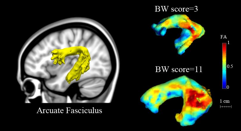
Previous studies have shown that in adults with poor reading skills, this structure, known as the arcuate fasciculus, is smaller and less organized than in adults who read normally. However, it was unknown if these differences cause reading difficulties or result from lack of reading experience.
“We were very interested in looking at children prior to reading instruction and whether you would see these kinds of differences,” says John Gabrieli, the Grover M. Hermann Professor of Health Sciences and Technology, professor of brain and cognitive sciences and a member of MIT’s McGovern Institute for Brain Research.
Gabrieli and Nadine Gaab, an assistant professor of pediatrics at Boston Children’s Hospital, are the senior authors of a paper describing the results in the Aug. 14 issue of the Journal of Neuroscience. Lead authors of the paper are MIT postdocs Zeynep Saygin and Elizabeth Norton.
The path to reading
The new study is part of a larger effort involving approximately 1,000 children at schools throughout Massachusetts and Rhode Island. At the beginning of kindergarten, children whose parents give permission to participate are assessed for pre-reading skills, such as being able to put words together from sounds.
“From that, we’re able to provide — at the beginning of kindergarten — a snapshot of how that child’s pre-reading abilities look relative to others in their classroom or other peers, which is a real benefit to the child’s parents and teachers,” Norton says.
The researchers then invite a subset of the children to come to MIT for brain imaging. The Journal of Neuroscience study included 40 children who had their brains scanned using a technique known as diffusion-weighted imaging, which is based on magnetic resonance imaging (MRI).
This type of imaging reveals the size and organization of the brain’s white matter — bundles of nerves that carry information between brain regions. The researchers focused on three white-matter tracts associated with reading skill, all located on the left side of the brain: the arcuate fasciculus, the inferior longitudinal fasciculus (ILF) and the superior longitudinal fasciculus (SLF).
When comparing the brain scans and the results of several different types of pre-reading tests, the researchers found a correlation between the size and organization of the arcuate fasciculus and performance on tests of phonological awareness — the ability to identify and manipulate the sounds of language.
Phonological awareness can be measured by testing how well children can segment sounds, identify them in isolation, and rearrange them to make new words. Strong phonological skills have previously been linked with ease of learning to read. “The first step in reading is to match the printed letters with the sounds of letters that you know exist in the world,” Norton says.
The researchers also tested the children on two other skills that have been shown to predict reading ability — rapid naming, which is the ability to name a series of familiar objects as quickly as you can, and the ability to name letters. They did not find any correlation between these skills and the size or organization of the white-matter structures scanned in this study.
Brian Wandell, director of Stanford University’s Center for Cognitive and Neurobiological Imaging, says the study is a valuable contribution to efforts to find biological markers that a child is likely to need extra help to learn to read.
“The work identifies a clear marker that predicts reading, and the marker is present at a very young age. Their results raise questions about the biological basis of the marker and provides scientists with excellent new targets for study,” says Wandell, who was not part of the research team.
Early intervention
The left arcuate fasciculus connects Broca’s area, which is involved in speech production, and Wernicke’s area, which is involved in understanding written and spoken language. A larger and more organized arcuate fasciculus could aid in communication between those two regions, the researchers say.
Gabrieli points out that the structural differences found in the study don’t necessarily reflect genetic differences; environmental influences could also be involved. “At the moment when the children arrive at kindergarten, which is approximately when we scan them, we don’t know what factors lead to these brain differences,” he says.
The researchers plan to follow three waves of children as they progress to second grade and evaluate whether the brain measures they have identified predict poor reading skills.
“We don’t know yet how it plays out over time, and that’s the big question: Can we, through a combination of behavioral and brain measures, get a lot more accurate at seeing who will become a dyslexic child, with the hope that that would motivate aggressive interventions that would help these children right from the start, instead of waiting for them to fail?” Gabrieli says.
For at least some dyslexic children, offering extra training in phonological skills can help them improve their reading skills later on, studies have shown.
The research was funded by the National Institutes of Health, the Poitras Center for Affective Disorders Research, the Ellison Medical Foundation and the Halis Family Foundation.
The study, done with researchers at Boston Children’s Hospital, found a correlation between poor pre-reading skills in kindergartners and the size of a brain structure that connects two language-processing areas.
Previous studies have shown that in adults with poor reading skills, this structure, known as the arcuate fasciculus, is smaller and less organized than in adults who read normally. However, it was unknown if these differences cause reading difficulties or result from lack of reading experience.
“We were very interested in looking at children prior to reading instruction and whether you would see these kinds of differences,” says John Gabrieli, the Grover M. Hermann Professor of Health Sciences and Technology, professor of brain and cognitive sciences and a member of MIT’s McGovern Institute for Brain Research.
Gabrieli and Nadine Gaab, an assistant professor of pediatrics at Boston Children’s Hospital, are the senior authors of a paper describing the results in the Aug. 14 issue of the Journal of Neuroscience. Lead authors of the paper are MIT postdocs Zeynep Saygin and Elizabeth Norton.
The path to reading
The new study is part of a larger effort involving approximately 1,000 children at schools throughout Massachusetts and Rhode Island. At the beginning of kindergarten, children whose parents give permission to participate are assessed for pre-reading skills, such as being able to put words together from sounds.
“From that, we’re able to provide — at the beginning of kindergarten — a snapshot of how that child’s pre-reading abilities look relative to others in their classroom or other peers, which is a real benefit to the child’s parents and teachers,” Norton says.
The researchers then invite a subset of the children to come to MIT for brain imaging. The Journal of Neuroscience study included 40 children who had their brains scanned using a technique known as diffusion-weighted imaging, which is based on magnetic resonance imaging (MRI).
This type of imaging reveals the size and organization of the brain’s white matter — bundles of nerves that carry information between brain regions. The researchers focused on three white-matter tracts associated with reading skill, all located on the left side of the brain: the arcuate fasciculus, the inferior longitudinal fasciculus (ILF) and the superior longitudinal fasciculus (SLF).
When comparing the brain scans and the results of several different types of pre-reading tests, the researchers found a correlation between the size and organization of the arcuate fasciculus and performance on tests of phonological awareness — the ability to identify and manipulate the sounds of language.
Phonological awareness can be measured by testing how well children can segment sounds, identify them in isolation, and rearrange them to make new words. Strong phonological skills have previously been linked with ease of learning to read. “The first step in reading is to match the printed letters with the sounds of letters that you know exist in the world,” Norton says.
The researchers also tested the children on two other skills that have been shown to predict reading ability — rapid naming, which is the ability to name a series of familiar objects as quickly as you can, and the ability to name letters. They did not find any correlation between these skills and the size or organization of the white-matter structures scanned in this study.
Brian Wandell, director of Stanford University’s Center for Cognitive and Neurobiological Imaging, says the study is a valuable contribution to efforts to find biological markers that a child is likely to need extra help to learn to read.
“The work identifies a clear marker that predicts reading, and the marker is present at a very young age. Their results raise questions about the biological basis of the marker and provides scientists with excellent new targets for study,” says Wandell, who was not part of the research team.
Early intervention
The left arcuate fasciculus connects Broca’s area, which is involved in speech production, and Wernicke’s area, which is involved in understanding written and spoken language. A larger and more organized arcuate fasciculus could aid in communication between those two regions, the researchers say.
Gabrieli points out that the structural differences found in the study don’t necessarily reflect genetic differences; environmental influences could also be involved. “At the moment when the children arrive at kindergarten, which is approximately when we scan them, we don’t know what factors lead to these brain differences,” he says.
The researchers plan to follow three waves of children as they progress to second grade and evaluate whether the brain measures they have identified predict poor reading skills.
“We don’t know yet how it plays out over time, and that’s the big question: Can we, through a combination of behavioral and brain measures, get a lot more accurate at seeing who will become a dyslexic child, with the hope that that would motivate aggressive interventions that would help these children right from the start, instead of waiting for them to fail?” Gabrieli says.
For at least some dyslexic children, offering extra training in phonological skills can help them improve their reading skills later on, studies have shown.
The research was funded by the National Institutes of Health, the Poitras Center for Affective Disorders Research, the Ellison Medical Foundation and the Halis Family Foundation.


