Skip to content ↓

Macular degeneration accompanied by significant brain reorganization

Press Inquiries

Press Contact:

Patti Richards
Phone: 617-253-2700
MIT News Office

Media Download

Images of human brains, based directly on data from an MRI machine, show the regions of the brain that are activated by peripheral visual stimuli in a person with normal vision and in a person with macular degeneration. People with macular degeneration lack central vision and must use peripheral vision only. The area at the back of the brain (marked by the white ovals) that normally responds only ...
Download Image
Caption: Images of human brains, based directly on data from an MRI machine, show the regions of the brain that are activated by peripheral visual stimuli in a person with normal vision and in a person with macular degeneration. People with macular degeneration lack central vision and must use peripheral vision only. The area at the back of the brain (marked by the white ovals) that normally responds only to stimuli in central vision, was strongly activated in the person with macular degeneration but not in the subject with normal vision. This shows that there has been reorganization of visual processing in the brain with macular degeneration.
Credits: Image / Chris Baker

*Terms of Use:

Images for download on the MIT News office website are made available to non-commercial entities, press and the general public under a Creative Commons Attribution Non-Commercial No Derivatives license. You may not alter the images provided, other than to crop them to size. A credit line must be used when reproducing images; if one is not provided below, credit the images to "MIT."

Close
Images of human brains, based directly on data from an MRI machine, show the regions of the brain that are activated by peripheral visual stimuli in a person with normal vision and in a person with macular degeneration. People with macular degeneration lack central vision and must use peripheral vision only. The area at the back of the brain (marked by the white ovals) that normally responds only ...
Caption:
Images of human brains, based directly on data from an MRI machine, show the regions of the brain that are activated by peripheral visual stimuli in a person with normal vision and in a person with macular degeneration. People with macular degeneration lack central vision and must use peripheral vision only. The area at the back of the brain (marked by the white ovals) that normally responds only to stimuli in central vision, was strongly activated in the person with macular degeneration but not in the subject with normal vision. This shows that there has been reorganization of visual processing in the brain with macular degeneration.
Credits:
Image / Chris Baker

The McGovern Institute for Brain Research at MIT, a leading research and teaching institute committed to advancing understanding of the human mind and communications, announced today that scientists have discovered the first evidence that brain reorganization occurs in people suffering from the progressive visual disorder macular degeneration.

The study, "Reorganization of Visual Processing in Macular Degeneration," was published in the Jan. 19 issue of The Journal of Neuroscience by MIT postdoctoral associate Chris Baker and Professor Nancy Kanwisher, in collaboration with Professor Eli Peli of the Schepens Eye Research Institute at Harvard Medical School.

Macular degeneration is the leading cause of blindness in the developed world, affecting more than 1.7 million people in the United States, and many millions of people worldwide. In macular degeneration, the center of the retina is damaged and sight is limited to peripheral vision. People suffering from this disease have blurry vision, which often causes severe difficulties with everyday tasks such as reading, driving and recognizing people.

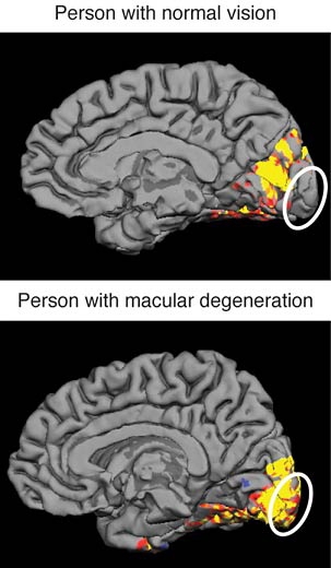
"Our major finding is that the part of the brain that processes only central retinal visual information in people with normal sight reorganizes itself in people with macular degeneration to help process peripheral visual information," said Baker.

Using advanced brain imaging techniques and state-of-the-art retinal mapping techniques, the researchers monitored which parts of the brain process visual signals in people with macular degeneration compared to people with normal vision.

"While our findings do not have immediate clinical application, the fact that a larger region of the cortex is recruited for peripheral vision in people with macular degeneration is encouraging, and suggests that it may be possible to develop new rehabilitation strategies that exploit this increased cortical involvement to partially compensate for loss of retinal function," said Kanwisher.

The researchers plan to explore whether the brain reorganization enables people with the disease to see better with peripheral vision than other people, and to identify the conditions that lead to these changes in the brain.

A version of this article appeared in MIT Tech Talk on January 26, 2005 (download PDF).

Related Links

Related Topics

More MIT News